Mehta limited his evaluation of FDG-PET (F-18 fluoro-2-deoxy-D-glucose) to nodal disease to facilitate its comparison with bronchoscopy. As he clicked through slides showing typically low-resolution PET scans of the thorax, there were hints that the modality wasn't his favorite.
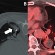
"This is a beautiful PET scan -- and a negative PET scan according to my nuclear radiologist. I think this is the reason why they sometimes call nuclear medicine 'unclear medicine.'"
From the literature, Mehta chose a prospective study that assessed PET's accuracy in detecting bronchogenic carcinoma. The double-blinded trial (reviewed by both a CT radiologist and a nuclear medicine physician blinded to clinical results) looked at 107 patients with abnormal chest x-rays. All 82 patients with increased FDG uptake had primary bronchogenic carcinoma. Sixteen of the patients with mediastinal metastases had increased localized FDG uptake, and 16 did not (American Journal or Respiratory and Critical Care Medicine, January 1996, Vol.153:1, pp.417-421).
"Twenty-five of the 107 patients had diseases unrelated to bronchogenic carcinoma," Mehta said. "Of course [PET] was positive in all 82 patients with lung cancer, but it was also positive in patients with sarcoidosis, coccidioidomycosis, microbacterial infection, and other inflammatory conditions involving the mediastinum. Even though it was sensitive, it was not very specific for the diagnosis of bronchogenic carcinoma. So individuals visiting from tuberculosis-endemic areas need to keep this information in mind."
Another study compared PET results to surgery in 30 patients with non-small cell lung cancer. The scan was correctly positive in seven patients with mediastinal involvement, but falsely negative in two. Overall, PET's positive predictive value was 96%, and the negative predictive value was 97%, Mehta said (AJRCCM, December 1995, Vol.152:6, Pt 1, pp.2090-2096).
The conference buzzed with talk of PET's high negative predictive value. Word was that a negative PET result is accurate enough to preclude invasive follow-up, but it's simply not true, Mehta said.
"Negative predictive value is prevalence-dependent. If there is zero prevalence of the disease, any test will have 100% negative predictive value," Mehta said. PET had other shortcomings too.
"A PET scan could be falsely positive in patients with tuberculoma, histoplasmoma, hamartoma, and anthracosilicosis," he said. It could be false-negative in patients with bronchoalveolar cell carcinoma, thyroid cancer, liposarcoma, and fibroma. Infection, acute inflammation, recent surgical wounds, and muscle hypermetabolism can give you false-positive results.... If there is a hilar lymph node next to a lesion, the PET scan could be negative."
Low-grade malignancy, hyperglycemia, and adjacent high metabolic focus also prompt FDG uptake. And there is no consensus on the quantitative analysis of PET scans, he said.
"You cannot let a tuberculosis patient not go for surgery, or let a histoplasmosis patient not go for surgery, and say that because your mediastinum is positive on a PET scan, you don't go for surgery," he said.
Because specificity is more important than sensitivity, and because PET remains expensive, limited in availability, and prone to false readings, it is not an adequate staging tool for bronchogenic carcinoma. Although the new PET/CT hybrid scanners could eventually have an impact, the technology remains unproven, Mehta said.
But PET comes with a bonus in its ability to find unsuspected distant metastasis, he said. For example, in a study of 99 non-small cell lung cancer patients, PET found 11 such metastases (Annals of Thoracic Surgery, December 1995, Vol. 60:6, pp.1573-1581).
In addition, PET imaging can be useful early in a workup, or when there is a suspected occult distal mass due to unexplained clinical symptoms such as weight loss or anemia, he said. It can help determine which CT-imaged lesions are benign and which are malignant. It can help select patients for neoadjuvant therapy. And it can be useful in patients who must avoid surgery, such as the elderly, or in those with coronary artery disease.

