German researchers have found that performing an MR angiography (MRA) scan for planning prior to uterine artery embolization (UAE) can cut both radiation and contrast dose in half, according to a study published in the June issue of Radiology.
UAE is an interventional radiology procedure that has proved to be a viable alternative to hysterectomy and myomectomy for women with uterine fibroids. But UAE is typically performed under fluoroscopy guidance, and radiation from the modality can be an issue for the very patients who are best suited to take advantage of the procedure: women of childbearing age.
Dr. Nagy Naguib and colleagues at the Johann Wolfgang Goethe University in Frankfurt, Germany, used 3D contrast-enhanced MRI angiography before UAE to predict the best angle for the angiography system's x-ray tube for imaging the uterine artery (Radiology, June 2009, Vol. 251:3, pp. 1-8). Choosing the right tube angle prior to the UAE procedure could eliminate the need to reposition the system during the exam, potentially reducing radiation dose.
"Unlike other oncology interventional procedures, like chemoembolization for malignant tumors, UAE is done for patients with benign disease, in their childbearing years," Naguib told AuntMinnie.com. "We're working near the ovaries, one of the most radiation-sensitive organs in the body, so it's crucial to use minimum radiation dose during intervention in such patients."
Forty women were included in the study: 20 sample patients who received 3D contrast-enhanced MRIs before UAE and 20 control patients who had undergone UAE without having MRI before the procedure. A single interventionist performed all the UAE procedures.
Contrast-enhanced MR angiography exams were performed with a 1.5-tesla MRI system (Magnetom Avanto, Siemens Healthcare, Erlangen, Germany). Two radiologists assessed images, performing all the 3D reconstructions together in consensus. The radiologists suggested two x-ray tube angles for each patient, one for imaging the left uterine artery origin and one for imaging the right uterine artery origin.
These possible angles were then provided to the interventional radiologist, who was asked to use them to better visualize the uterine artery origin during the procedure, but was blinded to the aims of the study (to reduce radiation and contrast medium dose).
UAE was successful in all 40 patients using a conventional angiography system (Axiom Artis, Siemens). After all the procedures had been performed, Naguib's team compared radiation dose, fluoroscopy time, and contrast medium volume between the sample group and the control group.
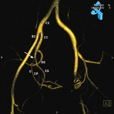 |
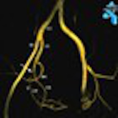
| Images in a 44-year-old woman. Above, 3D-reconstructed contrast-enhanced MR angiographic image from the pre-embolization study. The model was rotated to a 24° left obliquity for demonstration of the origin (arrow) of the right uterine artery (U) (routine oblique view). Upper right corner of the image shows the corresponding C-arm position and the obliquity angle (24° left oblique). Below, digital subtraction angiography (DSA) image in the same patient shows the corresponding angiographic projection and clear demonstration of the origin (arrow) of the right uterine artery (U). Additional arteries visualized include the common iliac (CI), external iliac (EI), inferior gluteal (IG), internal iliac (II), internal pudendal (IP), obturator (O), superior gluteal (SG), and superior vesical (SV). Both images courtesy of the Radiological Society of North America. |
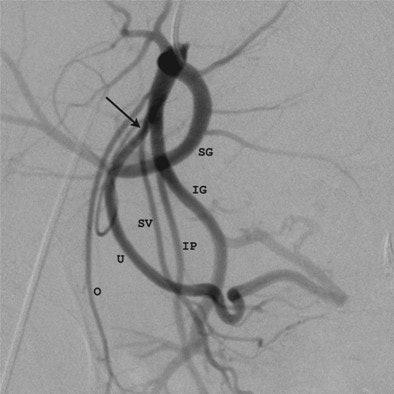 |
Naguib found that predicting the best tube angle for UAE using 3D contrast-enhanced MRI angiography helped.
|
"Due to the complex anatomy of the pelvic arteries, it's not possible to make a standard angle of obliquity for all women," Naguib said. "But our study shows that it is possible to predict the angle using 3D reconstructed MR angiography before performing UAE, so we don't have to find the correct angle using radiation."
By Kate Madden Yee
AuntMinnie.com staff writer
May 28, 2009
Related Reading
Hysterectomy most cost-effective fibroid treatment, March 20, 2009
Both UAE, hysterectomy improve quality of life, February 20, 2008
Copyright © 2009 AuntMinnie.com

