TORONTO - FDG-PET can play a useful role in the imaging of small-cell lung cancer (SCLC), according to two presentations Monday at the Society of Nuclear Medicine meeting.
Although FDG-PET has been readily deployed for non-small cell lung cancer imaging applications, the modality has not yet been proven of value in SCLC, which makes up approximately 20% of all lung cancers. But with the critical importance of initial staging and early detection required of SCLC, FDG-PET could fill an important niche, researchers said.
"[FDG-PET's] high sensitivity in SCLC may be very useful in initial staging in untreated patients," said Dr. Neeta Pandit-Taksar of Memorial Sloan-Kettering Cancer Center in New York City. "It also has a significant role in follow up by providing metabolic information in treated patients and may help in guiding therapy."
To evaluate FDG-PET in staging and assessing treatment response and cancer recurrence in SCLC, a Memorial Sloan-Kettering study team retrospectively reviewed 51 patients (67 scans) consisting of both treated (42) and untreated (9) cases. The imaging findings were correlated with pathology, CT/MRI, and clinical outcomes to determine sensitivity and specificity.
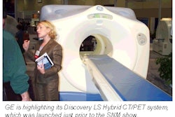
PET scanning was performed using an Advance PET scanner (GE Medical Systems, Waukesha, WI) following a six-hour fasting period and injection of 10 mCi of FDG. A pair of nuclear medicine physicians reviewed all cases; standardized uptake values (SUV) were calculated.
Overall, PET turned in a sensitivity of 90.2% with a specificity of 68.8%. In staging untreated cases, the scan findings concurred with final patient staging, Pandit-Taksar said. Mean SUVmax range in malignant cases was 7.6, while benign cases averaged 3.7. Both SUVmax (p=0.0075) and average SUV (p=0.0223) had significant negative association with survival, Pandit-Taskar said.
"SUVmax and positive PET study (results) provided prognostic information," she said.
Similar success with FDG-PET was reported in separate research at the department of nuclear medicine at Albert Einstein College of Medicine and Montefiore Medical Center in New York City. The Albert Einstein team retrospectively reviewed 15 patients with proven SCLC. Among the 15 patients, 3 were newly diagnosed and 12 had received chemotherapy and/or radiation.
Five patients (three newly diagnosed and two post-therapy) underwent surgery after the PET scan, and 14 received chemotherapy/radiation. One patient who was not treated with chemotherapy/radiation had surgery only.
All patients received CT scans prior to PET, with clinical follow up two months or more after PET. Following four hours of fasting, the whole-body PET scans were acquired approximately 50 minutes after injection of 3.4-4.15 mCi of FDG. The study team used a C-PET plus scanner (ADAC Laboratories, Milpitas, CA).
Among the 12 patients treated prior to PET, two were found to have solitary pulmonary nodules with FDG uptake on PET, said Dr. Deshan Zhao. Subsequent surgical resection and pathology revealed one true positive and one false positive case(post-radiation pneumonitis).
Six of the 12 patients had extrapulmonary metastases and/or large intense hilar or pulmonary uptake on PET, while four of the 12 had no evidence of abnormal FDG uptake. Three cases with newly diagnosed SCLC were all true positives on PET, confirmed by surgery, Zhao said. The study team attributed a false negative on the CT scan to post-radiotherapy fibrosis in the lungs.
The researchers concluded that whole-body FDG-PET is useful in evaluating the effect of treatment in SCLC patients, and can also provide the basis for determining the best treatment modality during the early stages of the cancer.
"FDG-PET can be helpful in selecting patients with SCLC who are candidates for surgical resection," Zhao said.
By Erik L. RidleyAuntMinnie.com staff writer
June 26, 2001
Click here to post your comments about this story. Please include the headline of the article in your message.
Copyright © 2001 AuntMinnie.com


