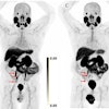
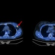
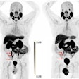
Diagnosing Alzheimer’s disease visually on amyloid PET scans has similar results to quantitative analysis methods used in research, according to a study published July 28 in JAMA Neurology.
The results are from an analysis of a large dataset derived from 602 clinical readers and 294 imaging facilities, including small community centers, and it supports the validity of amyloid PET visual reads, noted lead author Ehud Zeltzer, MD, of the University of California, San Francisco, and colleagues.
“These findings support the reliability of community [amyloid] PET scans as acquired and visually read in the real world,” the group wrote.
The use of amyloid PET scans in clinical practice is expected to increase with the expansion of Medicare coverage and the approval of amyloid-targeting drugs, which require positive PET scans for treatment eligibility, the authors explained. Previous studies validating the reliability of visual reads have used standardized approaches and experienced readers and, thus, require replication under the much less controlled environment of real-world clinical practice, they noted.
Thus, to assess the accuracy of clinical reads, the researchers compared visual interpretations of 10,350 amyloid-PET scans performed in real-world clinical settings to scan interpretations based on the “Centiloid scale,” a computerized technique that quantifies 3D pixel (voxel) volumes of amyloid deposits. PET scans were performed using one of three U.S.-approved amyloid PET radiotracers, with each scan classified as either positive or negative for cortical beta-amyloid based on the two methods.
According to the analysis, out of 10,350 scans, a total of 6,332 (61%) were visually read as positive, and 6,121 (59%) were quantitatively positive. The agreement between visual reads and quantitative classification was 86.3% (Cohen κ = 0.72), the researchers reported.
In addition, a total of 5,519 (53%) scans were both positive visually and quantitatively, and 3,416 (33%) were negative by both methods; 813 (8%) were positive visually and negative quantitatively, and 602 (6%) were negative visually and positive quantitatively.
“In this cross-sectional quality improvement study of a large heterogeneous dataset of real-world [beta amyloid]-PET scans, centrally extracted Centiloids showed a high concordance with local visual reads and comparable values and distributions with those reported in observational research studies and clinical trials,” the researchers wrote.
Ultimately, the study demonstrates the feasibility of Centiloid quantification in community-acquired amyloid PET scans, with this technique approved in Europe as an optional adjunct to visual assessments, the researchers noted.
“Our uniquely large imaging dataset, derived from many clinical facilities, including small community centers, from across the U.S., greatly strengthens the generalizability of our results,” the group concluded.
The full study is available here.


