The discovery of a thrombus within an aneurysm on an FDG-PET/CT scan can alter treatment for cancer patients and eliminate potentially major consequences, according to a study published online August 4 by the Journal of Nuclear Medicine.
Researchers at Saint Louis University in Missouri found 16 (1.7%) aneurysms in a retrospective review of 926 patients, with 15 aneurysms occurring in the abdominal aorta and one aneurysm in a patient's internal jugular vein. The investigation further discovered a thrombus in seven (44%) of the 16 aneurysm cases.
"Basically, one out of every 50 cancer PET/CT studies will show an aneurysm, and then half of them will have a thrombus," study co-author Dr. Medhat Osman, a professor of radiology and the director of nuclear medicine and PET/CT at St. Louis University, told AuntMinnie.com. "Even though it is a small number, we actually believe that the real numbers could turn out to be higher, because we know that we may be missing a small thrombus due to the spatial resolution of the PET scanner."
As noted in the study led by Dr. Razi Muzaffar, an internal medicine physician at the university, the elderly and patients receiving chemotherapy or radiation therapy typically undergo a PET/CT scan. This patient population is also at a greater risk of developing atherosclerotic cardiovascular disease.
AAA incidence
Previous studies have found that men between the ages of 45 and 54 have a 1.3% incidence of having an abdominal aortic aneurysm (AAA). That rate increases to 12.5% when men reach the ages of 75 to 84. Among women, the incidence ranges from 0% in the 45- to 54-year-old group and a 5.2% rate when they reach 75 to 84 years old.
However, as Osman and colleagues noted, the incidence of abdominal aortic aneurysm among cancer patients is not well-known.
Researchers reviewed 1,540 consecutive FDG-PET/CT studies (Gemini, Philips Healthcare) from 926 patients who were scanned due to known or suspected cancer.
A total of 16 aneurysms (1.7%) were found among the 926 cases, with aneurysms discovered in 12 men and four women. The 16 patients had a mean age of 72.6 years. Fifteen patients had an aneurysm in the abdominal aorta, while one patient had an aneurysm in the internal jugular vein.
Thrombus occurrence
Among the 16 patients with aneurysms, FDG-PET/CT suggested a thrombus in seven cases (44%), which was later confirmed on contrast-enhanced CT. The mean diameter of the aneurysms was 4.8 cm, ranging from 1.7 to 7.6 cm. The mean maximum standardized uptake value (SUV) of vessels with a thrombus was 1.9, ranging from 1.5 to 2.2.
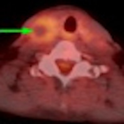
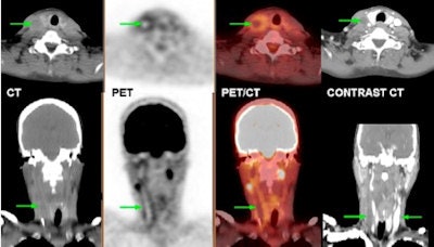
In the case of the patient with the occluded internal jugular vein, Osman said physicians intervened immediately and treated the condition with anticoagulation therapy. If the blockage had not been discovered, the result could have been fatal. "If it ruptures, it has a 90% mortality [rate]," he added.
 |
| A 57-year-old-woman with fully occluded left internal jugular vein. Unenhanced CT shows subtly hypodense left internal jugular vein. PET shows increased FDG uptake along course of vessel walls, with relative photopenia throughout lumen. Findings suggest accumulation of inflammatory and immune cells in vessel wall with fully occluded lumen. Contrast-enhanced CT confirmed findings and showed no enhancement of left internal jugular vein, as compared with right. Image courtesy of the Journal of Nuclear Medicine. |
This study is the first such research to determine the incidence of aneurysm in a population of cancer patients who received FDG-PET/CT, according to the authors.
"There is no data regarding cancer patients, even though we know that cancer is a risk for atherosclerotic disease by itself, as well as its therapies," Osman said. "Whether it is the disease or the cancer therapy that puts these patients at higher risk, there is no data at all about that. We were happily surprised that this is the first time that anyone reported the frequency of aneurysms in cancer patients."
In speaking with his radiation oncology colleagues at Saint Louis University, Osman said the study results have changed their approach to the management of cancer patients, whether it be adjusting therapies or radiation dose or the way the radiation is delivered. It could also alter patients' antithrombolytic or anticoagulant treatment.
"Clearly, [the results have] a clinical significance from the oncologist's point of view in the management of these cancer patients," Osman said. "This is where a large enough aneurysm and a large enough thrombus potentially may have a higher morbidity and mortality than the cancer itself."
As for potential study limitations, the authors noted that the CT scan was performed without contrast using a lower dose technique. "Given that thrombus formation is best visualized with contrast material, it is likely that the current study may have underestimated the frequency of thrombus formation within an aneurysm," they wrote.
Osman and colleagues currently are mulling whether to conduct a follow-up prospective study along those lines.
"We know that if we did not use contrast CT on everyone that we may be missing a small thrombus," he said. "It would be interesting if we do the CT with contrast to capture all of [the thrombi] and see how many -- if at all -- we are missing on the PET scan. The argument of using the added value of CT with contrast is not well-established in this setting."


