Wednesday, December 3 | 10:30 a.m.-10:40 a.m. | SSK18-01 | Room S505AB
FDG-PET/MRI and FDG-PET/CT topped diffusion-weighted MRI (DWI) in the diagnostic workup of lymphoma patients in this study from Swiss researchers at the University Hospital of Zurich.Lead author Dr. Marcelo Queiroz, from the department of medical imaging and nuclear medicine, and colleagues concluded that DWI-MRI is "less promising" in this clinical application because it was less accurate than FDG-PET/CT.
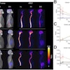
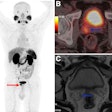
The aim of the study was to prospectively evaluate the accuracy of DWI compared with FDG-PET/MRI and FDG-PET/CT. The researchers used a trimodality protocol in which a patient undergoes a high-resolution whole-body MRI and then is transferred to the PET/CT using a shuttle board connecting the two systems to minimize patient movement and enable accurate image registration.
Queiroz and colleagues reviewed 83 FDG-PET/CT scans between April 2012 and January 2014, along with MRI data from whole-body DWI acquired with the trimodality configuration on 62 patients. They analyzed the results from PET/CT, PET/MRI, and DWI separately.
Imaging findings were confirmed by biopsy; follow-up imaging with CT, FDG-PET/CT, and/or FDG-PET/MRI; or clinical evaluation. Five scans were lost in follow-up.
In the review, FDG-PET/CT and FDG-PET/MRI detected disease in 29 patients. Both PET/CT and PET/MRI also correctly identified the stage of cancer, as well as all known 191 lesions.
Among the lymphoma patients, FDG-PET/CT and FDG-PET/MRI outperformed DWI in terms of sensitivity and specificity in the clinical setting. FDG-PET/MRI findings were in agreement with FDG-PET/CT for stage definition and disease detection.
"Our results reflect the challenges of an everyday clinical scenario confirming the anticipated equal performance of FDG-PET/CT and FDG-PET/MRI in the diagnostic workup of lymphomas," Queiroz wrote in an email to AuntMinnie.com.
DWI performed poorly compared with the other two modalities, regardless of which apparent diffusion coefficient cutoff was chosen to differentiate malignant and benign lesions, "leading to the conclusion that DWI does not provide additional relevant information in addition to FDG-PET/CT and/or FDG-PET/MRI," he added.


