Tuesday, December 1 | 3:10 p.m.-3:20 p.m. | SSJ02-02 | Room E450A
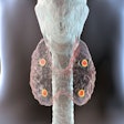
A Spanish company's positron emission mammography (PEM) system is drawing positive reviews from Dutch researchers, who will share their thoughts in this afternoon session.The Mammi-PET scanner is manufactured by OncoVision, a Valencia-based technology start-up created in 2003 by the University of Valencia and the Corpuscular Physics Institute. The PEM device is designed to create 3D images of the breast in a hanging position without the need for compression.
"Due to its high resolution, the Mammi-PET can visualize FDG-avid lesions as small as 2 mm," study presenter Dr. Suzana Teixeira, a doctoral candidate at the Netherlands Cancer Institute, told AuntMinnie.com.
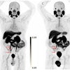
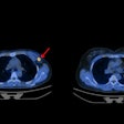
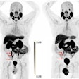
Teixeira and colleagues prospectively evaluated more than 200 women, each of whom had more than one histologically confirmed primary breast cancer lesion, between March 2011 and March 2014. The imaging protocol included an injection of 180 MBq to 240 MBq of FDG, followed by whole-body PET/CT.
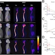
Mammi-PET achieved sensitivity just shy of 100% for lesions located within the device's range, and it detected almost all lesions smaller than 1 mm. Mammi-PET also found approximately 15 more lesions than PET/CT.
"The Mammi-PET was able to visualize the smallest breast cancer lesions in this series of 6 mm in diameter," Teixeira said. "Mammi-PET was also able to identify heterogeneity within primary breast cancer lesions, more so than is known for PET/CT."
She and her colleagues plan to advance their research by trying "to identify, at least visually, heterogeneous uptake within primary cancer lesions with the development of a Mammi-PET device with an integrated FDG-guided biopsy system," Teixeira said.
With PET-guided biopsies, tissue can be biopsied more specifically, which "opens the possibility to compare the histology of the most FDG-avid parts with those of less FDG uptake within the primary tumor," she added.


