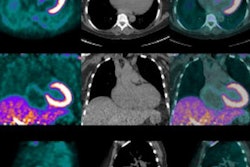
In the study, 30 patients with histologically confirmed neuroendocrine tumors underwent both hybrid scans with a single-injection protocol of Ga-68 DOTATOC and a fast PET/MRI sequence. Two readers independently analyzed both sets of images for lesion count, localization, malignancy, and conspicuity with histopathology and/or follow-up imaging as the reference standard.
The scans showed at least one malignant neuroendocrine tumor in 25 patients, with Ga-68 DOTATOC PET/MRI and Ga-68 DOTATOC PET/CT each correctly identifying 96% of these patients. Both modalities achieved similar results of 80% or greater for sensitivity, specificity, positive predictive value, negative predictive value, and diagnostic accuracy, and differences between the two techniques were not statistically significant (p = 0.38). In addition, maximum standardized uptake values were strongly correlated and did not differ significantly.
Lesion conspicuity, however, was better with Ga-68 DOTATOC PET/MRI (p < 0.01). While the study did not focus specifically on the clinical impact of whole-body staging differences between the two modalities, the researchers credited "superior soft-tissue contrast and benefits of diffusion-weighted MR imaging" for Ga-68 DOTATOC PET/MRI registering higher detectability of small liver metastases.
"As a majority of patients with high-grade neuroendocrine tumors develop liver metastases and the extent of metastastic spread is often crucial for differentiating a curative from a palliative situation, there might actually be a relevant clinical impact by using Ga-68 DOTATOC PET/MRI instead of Ga-68 DOTATOC PET/CT," wrote Dr. Lino Sawicki, from the department of diagnostic and interventional radiology at the University of Düsseldorf, in an email to AuntMinnie.com.
Sawicki and colleagues recommended that the findings be further investigated in future comparative studies.


