Dear Molecular Imaging Insider,
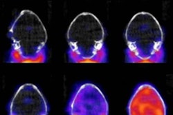
Quite often new healthcare techniques can produce events that seem like miracles. Take the case of French researchers who confirmed through PET and electroencephalography (EEG) the recovery of a patient who was in a vegetative state for 15 years after a car accident.
The group used vagus nerve stimulation to send electric pulses to the region of the brain associated with alertness and waking from sleep. After one month of treatment, the patient was able to obey verbal commands. That kind of remarkable progress certainly gives great hope for patients who are minimally conscious and their families.
Read more about the results and implications for vagus nerve stimulation in our Insider Exclusive.
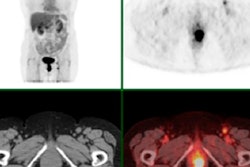
Another group of French researchers is promoting FDG-PET/CT for its ability to assess response to treatment and redirect therapy for patients with recurrent or progressive anal cancer. Using FDG uptake from the scans as an indication of metabolic response to chemoradiotherapy, radiologists were able to predict two-year progression-free survival. In cases of cancer recurrence, they also were able to direct patients toward other options such as surgery.
Artificial intelligence (AI) continues to make its mark in all elements of radiology. A research team from the University of Wisconsin in Madison has created an algorithm that can reliably classify benign and malignant bone lesions on F-18 sodium fluoride PET/CT images. AI eventually could make it possible to automate the time-consuming and subjective process for nuclear medicine physicians.
Researchers at Stanford University have developed a single-session PET/MR imaging protocol that reduces scan times for cancer patients and increases accuracy for evaluating chemotherapy-induced brain, heart, and bone abnormalities. The protocol reduced imaging time from 45 to 60 minutes to approximately 30 minutes or less per organ or region. The group also uncovered incidental findings that were not part of the initial targets.
Finally, U.S. Army researchers are contributing to the campaign to learn more about the progression of disease due to the Zika virus. The group is using PET scans of mice to study brain inflammation and visualize a variety of biological processes in live animal models.
Please stay in touch with the Molecular Imaging Community on a daily basis to be informed on the latest news and research from around the world.


