In a comparative imaging trial, fibroblast activation protein inhibitor (FAPI)-PET/CT outperformed FDG-PET/CT in detecting primary tumors in patients with head and neck cancer, according to a study published January 25 in the Journal of Nuclear Medicine.
The finding suggests that FAPI-PET/CT could serve as a sensitive, reliable, and reproducible imaging technique for these patients, in whom detection rates of primary tumors are low, noted Bingxin Gu, MD, of Fudan University in Shanghai, China, and colleagues.
“The low detection rate of primary tumors by current diagnostic techniques remains a major concern for patients with head and neck cancer of unknown primary,” the group wrote.
Head and neck cancer of “unknown primary” is a distinct group of highly heterogeneous malignancies, with patients typically presenting initially with enlarged cervical lymph nodes, the authors explained. Identifying where the cancer started in these patients using CT, MRI, nasopharyngoscopy, or laryngoscopy is a significant challenge, they wrote.
Studies have shown that F-18 FDG PET/CT molecular imaging can improve detection rates over these approaches based on visualizing glucose metabolism in tumor cells, yet nonspecific uptake of F-18 FDG radiotracer by inflamed tissue can lead to false-positive findings, they added.
Conversely, FAPI radiotracers such as gallium-68 (Ga-68) FAPI have been developed that bind to fibroblast cells in the supportive tissue of tumors, and these PET tracers have shown significant promise for detecting disease in previous studies.
In this study, the researchers tested the approach for the first time in head and neck cancer patients with tumors of unknown origin in a prospective clinical trial.
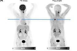
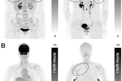
 PET/CT and MR images of a 72-year-old woman. (A) F-18 FDG-PET images (left and top) and PET/CT images (bottom) shown in coronal, sagittal, and axial views (from left to right), demonstrated a metastatic lymph node of the right neck with intensive metabolic activity (black arrow, SUVmax, 30.1) but presented no evidence for primary tumor. (B) Ga-68 FAPI-PET images (top and right) and PET/CT images (bottom), shown in axial, sagittal, and coronal views (from left to right), also detected metastatic lymph node with high Ga-68 FAPI tracer activity (black arrow, SUVmax, 16.3). There was intensive uptake of Ga-68 FAPI in palate (red arrow, SUVmax, 11.3). (C) T1-weighted, T2-weighted, and contrast-enhanced T1-weighted MRI also presented no evidence for primary tumor. Subsequent surgery confirmed mucoepidermoid carcinoma of palate. Image courtesy of the Journal of Nuclear Medicine.
The group enrolled 91 patients (18 women, 73 men; median age, 60 years old) with negative or equivocal findings of primary tumors by a comprehensive clinical examination and conventional imaging. Between June 2020 and September 2022, they performed F-18 FDG and (Ga-68) FAPI-PET/CT within one week, with the presence of primary tumors on the imaging sets then recorded by three experienced nuclear medicine physicians.
Primary tumors were detected in 46 (51%) patients after a thorough diagnostic work-up. According to the analysis, Ga-68 FAPI-PET/CT detected more primary lesions than F-18 FDG- PET/CT (46 vs. 17, p < 0.001) and showed better sensitivity, positive predictive value (PPV), and accuracy in locating primary tumors.
| Performance of Ga-68 FAPI-PET/CT vs. F-18 FDG-PET/CT | ||
|---|---|---|
| F-18 FDG | Ga-68 FAPI | |
| Sensitivity | 25% | 51% |
| PPV | 43% | 98% |
| Accuracy | 19% | 51% |
Moreover, Ga-68 FAPI-PET/CT led to improved treatment changes in 22 of 91 (24%) patients, the group added.
Ultimately, the study adds to promising results of Ga-68 FAPI PET/CT imaging in patients with various other head and neck cancers, such as nasopharyngeal carcinoma, oropharyngeal cancer, and salivary ductal carcinoma, the authors wrote.
Additional studies are warranted, especially given that the five-year overall survival of head and neck cancer patients with unknown primary tumors is dismal at 55%, they wrote.
“In the future, a multicenter trial needs to be performed to verify our results,” the group concluded.
The full article is available here.