Technetium-99m (Tc-99m) fibroblast activation protein inhibitor (FAPI) single-photon emission CT (SPECT) shows promise as a tool for assessing cellular and structural changes in the myocardium after heart attack, according to a study published July 14 in the Journal of Nuclear Cardiology.
Researchers at Capital Medical University in Beijing, China, found that the novel method revealed abnormal cellular activity five days after heart attack and detected so-called left ventricular (LV) remodeling after one year.
“The findings from this study have the potential to revolutionize post-[acute myocardial infarction] patient management by enabling early identification of those at risk for adverse LV remodeling,” noted first author Cuncun Hua, MD, and colleagues.
LV remodeling after heart attack is an adverse process in which cellular and structural changes occur in the myocardium, with the activation and proliferation of fibroblast cells playing a pivotal role. LV remodeling can lead to an increase in scar tissue in the heart, which is associated with heart failure.
While PET imaging with F-18- and gallium-68 (Ga-68)-labeled fibroblast activation protein inhibitor (FAPI) radiotracers has been established for evaluating this activity, the use of Tc-99m FAPI-SPECT has gained momentum due to its comparative cost-effectiveness, the authors wrote.
Thus, in this study, the researchers evaluated the approach further by testing it in 58 patients who experienced first-time ST-elevation myocardial infarction (STEMI), a type of heart attack caused by completely blocked coronary arteries. All patients (median age, 61 years old) underwent successful primary percutaneous coronary intervention (PCI) within 12 hours of symptom onset.
Patients underwent Tc-99m FAPI-SPECT five days after PCI and at 12-month follow-up visits. For comparison, the researchers recruited 33 healthy volunteers (average age, 32 years old) who also underwent Tc-99m FAPI-SPECT.
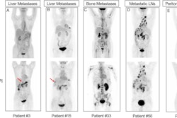
 Emergency percutaneous coronary intervention images, Tc-99m MIBI perfusion images and Tc-99m FAPI images of STEMI patients. In STEMI patients, there is a localized yet inhomogeneous uptake of Tc-99m FAPI. Notably, the extent of Tc-99m FAPI uptake significantly surpasses the area of perfusion defects. Despite similarities in coronary artery lesions (culprit vessels identified as the left anterior descending artery in image A and the right coronary artery in image B, white arrows), and comparable myocardial perfusion impairment, patients with larger Tc-99m FAPI uptake areas exhibit ventricular remodeling. Image courtesy of the Journal of Nuclear Cardiology.
According to the findings, baseline SPECT scans in the STEMI patients showed uptake of Tc-99m FAPI radiotracer in the myocardium indicating perfusion defect areas, while the healthy controls showed no detectable uptake.
Thirty patients completed a 12-month echocardiographic follow-up, and LV remodeling was identified in 11 patients (36.67%). Out of these 30, 15 patients had 12-month follow-up SPECT scans, with 13 still exhibiting abnormal Tc-99m FAPI uptake, the researchers noted.
“Our research demonstrates that this Tc-99m FAPI imaging modality holds promise in visualizing fibroblast activation following [myocardial infarction] and evaluating fibrotic changes,” the researchers wrote.
Ultimately, the study represents a significant step toward improving risk stratification and management in post-acute myocardial infarction patients, the authors suggested. Integrating the technique into clinical practice could enhance patient care by enabling early, targeted interventions, as well as offering advantages over PET imaging, including cost-effectiveness and wider accessibility, they wrote.
Nonetheless, additional research is needed to validate the findings, the authors noted.
“Further research is needed to validate these findings and to explore the broader implications of Tc-99m FAPI imaging in cardiovascular medicine,” the researchers concluded.
Access to the full article is available here.