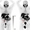
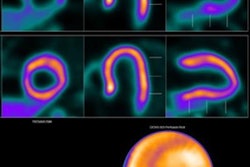
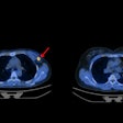
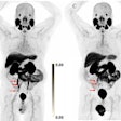
Cardiac PET/CT scans can effectively rule out moderate to severe cardiac allograft vasculopathy in heart transplant patients, according to an article published January 16 in the Journal of Nuclear Medicine.
The finding suggests that low-risk PET/CT scans can reduce the need for invasive coronary angiography screening in these patients, noted cardiologists at Columbia University in New York City.
“Recently, efforts have focused on developing noninvasive paradigms as an alternative to angiography,” wrote first author Nikil Prasad, MD, and colleagues.
Cardiac allograft vasculopathy (CAV) is a process unique to heart transplant recipients that causes impaired blood flow in both epicardial vessels and microvasculature, ultimately leading to allograft dysfunction, the researchers explained. Current standards for routine CAV screening recommend the use of invasive coronary angiography (ICA) to assess the epicardial vessels at regular intervals.
Yet yearly ICA exposes patients to a significant cumulative volume of iodinated contrast and poses unique risks to transplant patients, the group noted. Conversely, PET/CT myocardial perfusion imaging (MPI) scans are a proven low-risk and powerful method for identifying microvascular dysfunction.
In this study, the researchers analyzed whether PET/CT MPI can rule out whether patients may progress to CAV and thus reduce the need for ICA.
The work included 344 adult heart transplant patients who continued routine CAV screening and who also had at least one baseline ICA before and one subsequent ICA after PET/CT MPI scans. Patients were split into three groups based on the intervals between the ICA procedures: 450 to 750 days, 750 to 1,050 days, and 1,050 to 1,450 days. CAV was classified as absent (CAV 0), mild (CAV 1), moderate (CAV 2), or severe (CAV 3) according to current guidelines.
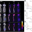
The analysis found that grade 0/1 CAV on PET/CT had a negative predictive value (NPV) of 93%, 95%, and 95% at each respective time point for developing moderate to severe CAV on subsequent ICA. Compared with PET CAV 0, a grade of CAV 2/3 on PET/CT was associated with a 2.9-fold increased risk of all-cause mortality (hazard ratio, 2.86).
In addition, in a sensitivity analysis of 135 patients with apparently stable CAV over successive ICA procedures, a grade of CAV 2/3 on PET/CT was associated with an increased risk of death or retransplantation (hazard ratio, 3.2), the group found.
“The frequency of invasive CAV screening may be reduced for patients with PET CAV 0/1, especially among those without subsequent rejection or development of donor-specific antibodies,” the researchers wrote.
The authors noted that their institution only recently switched its CAV screening protocol to include biennial PET/CT scans, which limited the mean follow-up time in the study to approximately five years. Nonetheless, the study validates PET/CT MPI as a diagnostic tool with prognostic value for the development of CAV between invasive angiographies, they wrote.
“Further investigation is required to better assess the external validity of these results across a more heterogeneous spectrum of disease,” the researchers concluded.
The full study is available here.


