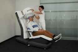
A Massachusetts company is hoping that renewed interest in functional brain imaging could help it find a market for a novel SPECT technology it developed decades ago, before the arrival of CT and MRI decimated the nuclear medicine brain imaging market.
The NeuroFocus scanner developed by NeuroPhysics of Devens, MA, produces higher resolution functional brain images than most PET scanners -- at the cost of a traditional gamma camera, according to NeuroPhysics founder Hugh Stoddart, the company's vice chairman and senior scientist. Stoddart hopes to retrieve some of the brain imaging market that nuclear medicine has lost over the past decade to CT and MRI.
Like other SPECT cameras, the vendor's technology is based on single-photon radiopharmaceuticals, but is unrelated to the multiheaded rotating gamma cameras commonly used for SPECT. NeuroPhysics calls its technology HDFET, for high-definition focused-emission tomography. The scanner uses point-focus collimators that the company calls "gamma lenses" in a technology related to focal point scanning biological microscopes.
Twelve long-bore lenses -- each coupled to its own sodium iodide crystal detector -- are arrayed at 30º intervals around the patient's head. The focal points lie inside the head. Following an injection of DuPont's Neurolite technetium-based radiopharmaceutical, the NeuroFocus scanner moves side to side at a right angle to the table, while the lenses move in and out, uniformly sampling a transaxial slice. Slices of the entire head are obtained by moving the patient bed in and out of the scanner bore.
Stoddart and his son, Hugh Stoddart, patented the NeuroFocus scanner in 1980, and were able to grandfather the unit with a 510(k) clearance when the Food and Drug Administration implemented the 510(k) regulatory framework. But the development of CT and MRI undercut the use of SPECT for brain imaging, Stoddart said.
"Since the nuclear medicine people were losing the head business, this machine was used primarily for brain research at Harvard, Columbia, Yale, and the Lawrence Berkeley National Laboratory (LBL)," Stoddart said. It was shown at the RSNA meeting just once, in 1993.
So the technology languished for 20 years until computers gained enough speed to make higher resolution possible and demand developed for such a machine. Today, an aging population is driving the functional brain imaging market with more brain diseases and disorders. And a growing pharmacopoeia to treat clinical brain indications is creating a need for functional brain imaging equipment to noninvasively monitor brain drug therapies.
Alzheimer's disease is a perfect example. HDFET's resolution is at least 3 mm, fine enough to see structures as well as function, Stoddart said. With biomarker imaging, this resolution allows neurologists to identify the striatum typical of Alzheimer's, something CT and MRI cannot do effectively at present.
Only PET, which is much more expensive and uses a fluorodeoxyglucose (FDG) radioisotope, can provide images with similar resolution. FDG has a half-life of less than two hours, however, while Neurolite's half-life is up to six hours. A typical dedicated PET camera costs more than $1 million; the NeuroPhysics brain scanner comes in at around $600,000.
The world-famous LBL is better known for developing nuclear weapons, but its Center for Functional Imaging has been using Stoddart's scanner for 15 years in gene therapy research on rhesus monkeys.
Center director Dr. Thomas Budinger is a nuclear medicine physician and functional imaging specialist. He says the company's SPECT scanner gives the highest resolution of any he has seen.
"In evaluating tomographic systems, we did some experiments on phantoms, and I became convinced the system Hugh Stoddart and his son had devised had promise," Budinger said. "How they had configured the system was more efficient than anyone could imagine. This scanner has sufficient resolution to allow us to see the efficacy of new approaches to treating Parkinson's disease."
The most important uses for the brain scanner -- and the areas where fMRI just can't compete -- are in post-stroke, Parkinson's, and Alzheimer's assessment; in measuring seratonin levels in depressive disease; and in studying the propensity for addiction, Budinger said.
To capitalize on the renewed interest in functional brain imaging, NeuroPhysics hopes to find a commercial partner with the necessary sales infrastructure as the fastest route to bringing the NeuroFocus scanner to market, according to NeuroPhysics CEO Dennis Caulfield. NeuroPhysics is in discussions with two potential partners.
Caulfield said the company is looking for "any major medical company with hitting power in the medical market."
You probably won't see NeuroPhysics at this year's Society of Nuclear Medicine meeting. In addition to the cost of putting on an exhibit, Stoddart and Caulfield do not believe there is much to be gained from marketing to nuclear medicine physicians. They plan to go straight to brain specialists.
"While this is a nuclear medicine machine, it's a neurologist or psychiatrist or even an oncologist who has the immediate need, because they have the patients," Caulfield said. "Nuclear medicine people would love to have it, but they are a service organization and don't have the horsepower to support (a purchase)."
By Robert BruceAuntMinnie.com contributing writer
March 13, 2001
Related Reading
PET and MRI race to detect early Alzheimer’s, January 22, 2001
FDG-PET clears up metabolic murkiness between dementia and Alzheimer's, November 7, 2000
Adding SPECT, MRI to Alzheimer's work-up costs more than it's worth, analysts say, October 11, 2000
Click here to post your comments about this story. Please include the headline of the article in your message.
Copyright © 2001 AuntMinnie.com


