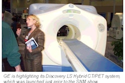
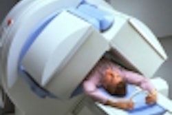
TORONTO - For evaluating patients with suspected primary lung cancer, nuclear medicine has come a long way, baby. On the road from gallium and V/Q to promising new radionuclides such as depreotide, to CT fusion techniques that could finally get radiotherapy planning right, Dr. Alan Waxman sees nuclear medicine's bright spots.
Waxman, a nuclear medicine specialist and co-chair of imaging at Cedars-Sinai Medical Center in Los Angeles, discussed new lung cancer imaging techniques in a series of Saturday sessions at the Society of Nuclear Medicine show on the state of the art in nuclear medicine.
"We all remember the gallium [GA-67] papers that came out 15-20 years ago...they actually have some good information, and it turns out that gallium was not terrible in looking at solitary pulmonary nodules [SPN] for metastases to the hilum and mediastinum" he said. "But [compared] with the new pharmaceuticals, recently thallium [T1-201] is showing its superiority, [Tc-99m] sestamibi actually showing a superiority over [both of] these, and now we're getting into areas such as Neotect [Tc-99m-labeled depreotide], which has been approved for SPN, and PET imaging, which is making a very bold move now after reimbursement has been established."
The essential areas in lung tumor assessment include detecting SPN, and then determining whether the tumor has spread to the hilum and mediastinum, and then looking for distant metastases, Waxman said. Many new tools and techniques are coming online to assist in this task, and the challenge is figuring out where they fit into the picture.
New crystals
Along with the well-established sodium iodide (NaI) and bismuth germanate (BGO) PET scanners, new high-end scanners are incorporating new LSO and GSO crystals that promise higher scanning resolution, both for SPN and for distal metastases. The new crystals may not make the pictures any prettier, Waxman said, but they promise to get patients in and out of the nuclear medicine department faster.
Even as resolution increases to the point where structures just a few millimeters in size can be seen, an important question is whether the scans are really seeing cancer, or perhaps granulomous disease in the hilar structures, for example. Explaining the hot spots can be especially daunting now that resolution has enabled visualization of suspicious structures too small to be seen on CT, a trend that is likely to continue, he said.
A big problem with CT is how suspected nodules are handled clinically. Large, growing nodules seen on CT are of course removed, but patients with indeterminate nodules -- small structures with rounded rather than spiculated borders -- are subjected to a "wait and watch" philosophy that can be unnerving.
"If you get CTs every three months, the patients go nuts waiting for the next CT. They 'know' they're going to die, and it's absolutely the end of their life; they have lung cancer -- even if they've never been a smoker."
At least incrementally, evaluation with PET or depreotide makes the job a little easier for both doctor and patient. Whatever lights up on the scans is alive -- not necessarily cancer -- but alive, Waxman said.
From there it gets trickier, but some researchers are now looking to quantify the nuclear medicine scan results by looking at the tumor-to-mediastinum ratios, or to the standard uptake values (SUV). Areas scoring below 2.5 are generally read as benign.
PET's accuracy
A recent meta-analysis published in the Journal of the American Medical Association (JAMA 2001 Feb 21;285(7):914-24) looked at a large number of PET studies that included at least 10 patients, at least five of whom had tumors that proved to be malignant. In a review of 1,474 focal lesions that were found, PET had a sensitivity and specificity of 96.8% and 77.8%, respectively. In contrast, CT is generally reported to have specificity for SPN in the 65%-70% range. A recent study by Falk and colleagues also found PET to be 22% more sensitive than CT for hilar and mediastinal staging, Waxman said.
An ongoing study at his own institution has evaluated more than 100 patients thus far, with very promising preliminary results. In 52 of the subjects, 32 were negative for CT and 20 were positive. In all, 13 of 32 negative CT patients were found to have cancer.
"CT totally blew it," Waxman said. "PET, on the other hand, picked up 10 of the 13 who were positive for cancer. If you look at the 19 negative pathology patients [19+13=32], 5 false positives were seen. The negative predictive value (NPV) for CT was 59%, the NVP for PET was 86%, keeping in line pretty much with what the literature is saying. That means that for every three patients taken to surgery, 2 of those patients will have true positives, and one will have a false positive. That's a very acceptable methodology to use to determine who goes to mediastinoscopy," he said.
Moreover, thoracic surgeons and pulmonologists at Waxman’s institution, who now order nuclear medicine scans in all suspected primary lung cancer patients, give PET results great sway in determining candidates for mediastinoscopy. At Cedars-Sinai, a negative CT is never used to eliminate mediastinoscopy, a procedure whose results are highly operator-dependent.
Nevertheless, Waxman advised caution with regard to interpreting PET results in light of its well-known proclivity for producing false-positive results in granulomous disease, tuberculosis, or other active infection, and even in heart disease. PET-imaged "tails" heading into the mediastinum are especially difficult to call, and may or may not be malignant, he said.
The big news, of course, is CT/PET fusion, which is calming the crosstown rivalry between the two modalities. Fusion's most important role in pulmonary medicine is in radiation therapy planning.
A variety of fusion techniques are available, including hardware- and software-based and fiducial and nonfiducial techniques that vary in complexity, he said. If fiduciary marks are used, same-day imaging with both modalities may be necessary to prevent patients from washing them off, he added.
For its part, Cedars-Sinai employs combined hardware systems for PET and CT to produce DICOM data, using both emission and transmission fusion techniques, Waxman said. Since emission fusion has been much more difficult to accomplish than transmission fusion, the physicians have developed a hybrid acquisition technique. They collect the transmission image of the PET scan, fuse it to the CT image, alter the transmission image to the same coordinates (the patient is not being moved) and then make the emission image reappear in the same area, Waxman said.
"The registration precision is plus or minus 5 mm tolerance, according to our radiation oncologist," he said, showing a fusion image marked for radiation therapy by both PET and CT. In an affirmation of fusion's value, it turns out the CT isocontour would have resulted in unnecessary irradiation of the aorta along with the tumor, because the difference between the structures was unremarkable on CT.
"It was striking for me to see that these two individuals, both very highly trained and highly intelligent, had very different fields that they were going to irradiate."
Already a study of 10 patients at his institution has showed an average radiation target volume change of 156 cc, or 71%, in 10 patients.
"Tumor volume irradiated was always increasing as they showed additional abnormalities in PET. We also don't have -- and we need -- long-term outcomes data to determine if we're doing any good. We're irradiating larger areas and giving a boost to some very specific sites that we're not seeing on CT. The question is [whether] we are extending life, can we improve the quality of life, and if so by how long?"
By Eric Barnes
AuntMinnie.com staff writer
June 25, 2001
Click here to post your comments about this story. Please include the headline of the article in your message.
Copyright © 2001 AuntMinnie.com


