LOS ANGELES - FDG-PET imaging has been very useful to clinicians trying to determine whether a pulmonary nodule is a benign or neoplastic lesion. For the most part, these studies have been performed at a single point in time. However, FDG uptake from inflammatory processes peaks earlier than in most malignant disorders, which can lead to false-positive readings.
"Single-time-point FDG-PET imaging is highly accurate in distinguishing the malignancy of pulmonary nodules, but significant false-positive results by some investigators remains a problem," said Dr. Marc Hickeson.
Hickeson and a team of fellow researchers from the Hospital of the University of Pennsylvania in Philadelphia outlined a protocol to improve the accuracy of pulmonary nodule FDG-PET imaging to attendees at the 49th Society of Nuclear Medicine annual meeting. The technique involves imaging patients at two time points versus traditional single-time-point PET.
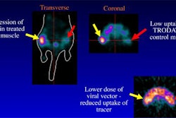
The group, through experiments on animal and cell models, determined that there is an increase in the intensity of malignancies over time due to the glucose metabolism of tumors. In contrast, they found that the intensity of benign lesions decreased or remained stable over the same period. On the basis of this research, the investigators conducted a retrospective study of 141 patients who underwent dual-time FDG-PET scans at their facility.
The patients all had lung lesions that were confirmed by pathology. The first PET scans were conducted 48-71 minutes after the injection of FDG. The second scans were performed 41-69 minutes after completion of the first scan. The region of interest was set by the researchers at 4 pixels over the highest uptake region.
A malignancy was determined by the team to be a 10% increase of intensity on the dual-point scans. An intensity increase of 9.9% or less was labeled as benign. The interpretation of the images was compared with final results based on surgical pathology, biopsy, repeated radiographic examinations, or clinical outcome, Hickeson said.
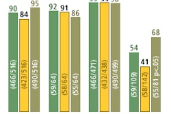
The team found that the sensitivity, specificity, and accuracy of single-point FDG-PET imaging for characterizing the pulmonary nodules was 88.3%, 83%, and 86.5%, respectively. The dual-point PET imaging had a sensitivity, specificity, and accuracy of 95.7%, 85.1%, and 92.2% respectively.
When the sensitivity of a test increases due to interpretation criteria, specificity and positive predictive values generally decline as a result, Hickeson said. However, his group’s data indicates that dual-point FDG-PET imaging increased the sensitivity without adversely affecting the specificity of the test.
"Although our work clearly demonstrates the superiority of dual versus single-time-point FDG-PET imaging in the assessment of pulmonary nodules, further studies will be required to determine the optimal imaging protocol," Hickeson said.
By Jonathan S. Batchelor
AuntMinnie.com staff writer
June 21, 2002
Copyright © 2002 AuntMinnie.com


