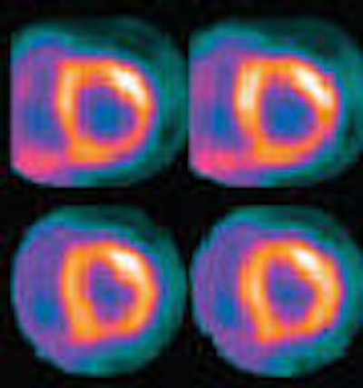
Finding and identifying disease through noninvasive imaging is a critical element of any exam, but excluding disease is also important for its role in a patient's prognosis and treatment. While molecular imaging for coronary artery disease (CAD) with SPECT or PET is accurate, diagnosis of multivessel CAD has proved difficult with these modalities.
A team of researchers from Brigham and Women's Hospital in Boston may have found a way to address the insufficiencies of multivessel CAD diagnosis by utilizing rubidium (Rb-82) PET/CT to determine a value for vasodilator left ventricular ejection fraction (LVEF) reserve. This value is then used in evaluating the magnitude of myocardium at risk and the extent of angiographic CAD.
"Unlike vasodilator SPECT with technetium agents in which stress gated images are obtained approximately 45 minutes after stress, Rb-82 PET enables measurement of left ventricular function during peak hyperemia," the authors wrote in a study published in the Journal of Nuclear Medicine (March 2007, Vol. 48:3, pp. 350-358).
The team studied 510 consecutive patients (mean age 61 years with females comprising 59% of the cohort) with suspected CAD who underwent gated rest and vasodilator stress Rb-82 PET/CT on a Discovery ST scanner (GE Healthcare, Chalfont St. Giles, U.K.). The study group was comprised of 44 control patients, 309 patients with normal myocardial perfusion imaging (MPI), and 157 patients with abnormal MPI.
"The CT portion of PET/CT in this analysis was used solely for correction of photon attenuation by the soft tissues and was not used for a calcium score or coronary angiography," the authors noted.
Gated myocardial image acquisition was begun 90-120 seconds after injection of 1,480-2,220 MBq of Rb-82 for the rest portion of the exam, and continued for five minutes. Vasodilator stress was induced with adenosine or dipyridamole infusion, a second dose of Rb-82 was administered, and gated images were acquired in the same manner as the resting images.
The scientists calculated the LVEF as well as the left ventricular end-diastolic volumes and end-systolic volumes using the Emory Cardiac Toolbox by Emory University Hospital in Atlanta. They noted that the LVEF reserve was reported as the absolute difference in ejection-fraction percentage. Two observers assessed the myocardial perfusion images using a 17-segment model.
 |
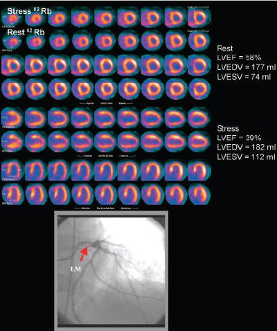
| Rest-stress myocardial perfusion PET/CT images in patient with severe angina demonstrate small region of moderate reversibility in middle and basal inferolateral walls. Gated study demonstrated significant decrease in LVEF during peak stress. Catheter coronary angiography demonstrated left dominant anatomy and severe left main coronary artery disease (bottom panel). Patient underwent coronary artery bypass surgery. LVEDV = left ventricular end-diastolic volume; LVESV = left ventricular end-systolic volume; LM = left main coronary artery. Reprinted with permission from "Value of Vasodilator Left Ventricular Ejection Fraction Reserve in Evaluation the Magnitude of Myocardium at Risk and the Extent of Angiographic Coronary Artery Disease: A 82Rb PET/CT Study;" Sharmila Dorbala, Divya Vangala, Uchechukwu Sampson, Atul Limaye, Raymond Kwong, and Marcelo F. Di Carli; March 2007, Journal of Nuclear Medicine. |
A total of 68 patients from the Rb-82 PET/CT group had coronary angiography within six months of their perfusion imaging exam and the researchers obtained cine angiograms with an Integris BH3000 angiographic system (Philips Medical Systems, Andover, MA). Severe CAD was determined visually as a stenosis diameter of 70% or greater for the left anterior descending, left circumflex, and right coronary arteries or their major branches, and 50% or greater for the left main coronary segment as described in the patients' clinical coronary angiogram reports, the authors reported.
"Of these 68 patients, 15 (22%) showed nonobstructive CAD, 23 (34%) single-vessel CAD, 13 (19%) two-vessel CAD, 9 (13%) three-vessel CAD, and eight (12%) left main disease," they wrote.
Using multiple logistic regression analysis, the scientists concluded that the only significant independent predictor of left main or three-vessel CAD was the LVEF reserve. They determined that for each unit increase in LVEF reserve, the odds of left main or three-vessel CAD decreased by 20%; for each unit decrease in LVEF reserve, the odds of left main or three-vessel CAD increased by 30%.
The researchers noted that obtaining an LVEF reserve parameter during a rest-stress Rb-82 PET exam is not difficult, and that the information it provides can exclude severe left main or three-vessel CAD with a high degree of certainty.
"In a large number of patients with suspected CAD, the current study results demonstrate that measures of LVEF reserve assessed during vasodilator stress Rb-82 PET can be used as an aid to perfusion imaging to better delineate the magnitude of jeopardized myocardium and more precisely define the extent of underlying anatomic CAD compared with perfusion data alone," they concluded.
By Jonathan S. Batchelor
AuntMinnie.com staff writer
March 30, 2007
Related Reading
Automated emission correction shows potential for cardiac PET/CT, March 9, 2007
PET identifies inflammation severity in carotid plaques, November 21, 2006
PET perfusion can predict cardiac events, March 15, 2006
PET, SPECT measures of LVEF have superior predictive value, March 13, 2006
Rubidium-based PET may finally get its due, February 23, 2006
Copyright © 2007 AuntMinnie.com


