A study from the University of Texas M. D. Anderson Cancer Center in Houston has found that FDG-PET/CT can help determine progression-free survival, overall survival, and tumor necrosis among patients with osteosarcoma.
The retrospective study focused on maximum standardized uptake value (SUVmax), total lesion glycolysis (TLG), and changes between SUVmax and TLG in patients' FDG-PET/CT scans before and after initial chemotherapy.
PET/CT "has been found to be extremely useful, particularly for follow-up of treatment for therapeutic effect in various types of cancer. It also has been investigated for prognostic values," said Dr. Colleen Costelloe, lead author and assistant professor of radiology, musculoskeletal section, at M. D. Anderson. "While treatment effects tend to be fairly well represented with the use of PET/CT, predictor of outcome at the outset is somewhat up in the air."
Even though osteosarcoma is the most common nonhematologic primary bone tumor, the cancer is not covered by Medicare reimbursement. Therefore, Costelloe noted, studies of osteosarcoma and PET/CT are still relatively few in number.
The study, published in the Journal of Nuclear Medicine (March 2009, Vol. 50:3, pp. 340-347), is the first paper to "meaningfully evaluate" TLG in patients with osteosarcoma, the authors wrote. "Unlike SUV, which is a measurement of metabolic activity per body weight, TLG evaluates metabolic activity throughout the volume of the tumor (SUVavg multiplied by tumor volume) above a minimum threshold designed to exclude background activity."
Patient population
The retrospective study included 19 men and 12 women, ranging in age from 9 to 65 years, at M. D. Anderson between January 1, 2002, and January 1, 2008. All 31 patients underwent FDG-PET/CT before and after chemotherapy, followed by tumor resection. Two radiologists experienced in PET/CT interpretation measured the SUVmax and TLG on all prechemotherapy and postchemotherapy scans.
PET, CT, and PET/CT fusion datasets were reviewed. With a volumetric region of interest centered on the lesion, the tumor osteoid in all lesions defined the margins of the tumor irrespective of decrease in FDG uptake after therapy. All tumors were more than 3 cm in size.
SUV and TLG
The SUV was defined as measured activity concentration (Bq/mL) multiplied by lean body mass (kg) divided by injected activity (Bq). TLG was defined as SUVavg times tumor volume, with a threshold of 45% SUVmax in the volume of interest.
Costelloe said it is important to note that researchers calculated SUVmax using lean body mass to eliminate bias in the results due to a cancer patient's fluctuating weight. "A cancer patient may have lost or gained weight between exams; total body weight will be affected by it," she added. "The bottom line is that the cut point value will not be exact, as at other institutions, but it is a good ballpark."
Integrated PET/CT systems (Discovery ST, STE, or RX from GE Healthcare, Chalfont St. Giles, U.K.) were used to acquire whole-body examinations from the vertex of the skull through the upper thighs, lower legs, or toes, depending on the location of the primary tumor.
All patients had fasted for a minimum of six hours, with a blood glucose level of 80-120 mg/dL before intravenous administration of FDG (555-740 MBq). An unenhanced CT scan was used for attenuation correction. Patient follow-up ranged from 152 to 1,623 days, with a median follow-up of 2.6 years.
Patient reviews
In the patient review, the researchers found a correlation between SUVmax and progression-free survival. Poor outcome was associated with higher values for both the prechemotherapeutic SUVmax and the postchemotherapeutic SUVmax. A prechemotherapeutic SUVmax of less than or equal to 15 g/mL was significant with progression in 15% of patients.
"With SUVmax, the computer will pick out the point of greatest metabolic uptake in that volume," Costelloe said. "For TLG, it is the SUV average above a certain threshold. It's a different analysis. The question is: Which method is a better predictor? This study showed that SUVmax does, indeed, seem to be the stronger predictor."
The data also revealed that a postchemotherapeutic SUVmax measurement of ≥ 5 g/mL was significantly associated with progression in 23% of patients above the cut point.
"Although there was no evidence that TLG values on the pre- or the postchemotherapeutic examinations were associated with progression-free survival," the researchers wrote, "an increase in TLG between the two examinations was associated with a shorter progression-free survival."
 |
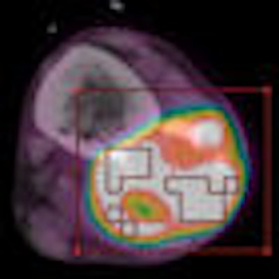
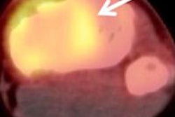
| The fused FDG-PET/CT images belong to a 25-year-old male patient with high-grade conventional osteosarcoma of distal fibular metaphysis. Coronal (A) and axial (B) images of the tumor before chemotherapy demonstrate SUVmax and TLG values of 10.8 g/mL and 159.5 g, respectively. Coronal (C) and axial (D) images after completion of chemotherapy show significant reduction in SUVmax to 2.3 g/mL and TLG to 85.8 g. Images courtesy of the Journal of Nuclear Medicine and M. D. Anderson Cancer Center. |
 |
The researchers also found that high SUVmax on postchemotherapeutic examinations was associated with increased mortality; patients with an SUVmax greater than or equal to the median of 3.3 g/mL had a higher likelihood of dying. SUVmax before chemotherapy was not associated with increased mortality.
Regarding tumor necrosis, good overall and progression-free survival was associated with tumor necrosis greater than 90%. Tumor necrosis greater than 90% was most strongly associated with a decrease in SUVmax.
The study "comes definitively on the side of PET/CT being able to predict survival, which means that one can theoretically modify the therapy ahead of time, instead of waiting for the tumor to not respond to typical therapies," Costelloe said.
The authors acknowledged that one study limitation was the relatively small sample and short follow-up of patients, but they added that the research is the "largest series evaluating neoadjuvant chemotherapy for osteosarcoma by PET." They recommended a longer patient follow-up period to better assess survival because osteosarcoma typically recurs within two to three years of surgery.
"We need larger numbers and we need prospective study design in order to verify PET/CT in general is a reliable indicator of outcome, upon which clinicians may wish to modify initial therapy, instead of waiting for an initial suboptimal result," Costelloe said. "That would truly require a large, randomized, prospective study to convince clinicians to do that. This study has planted the seed for justifying that kind of study."
By Wayne Forrest
AuntMinnie.com staff writer
March 12, 2009
Related Reading
PET/CT brings added value to pediatric oncology, February 27, 2004
FDG PET reveals osteosarcoma's response to chemotherapy, October 1, 1999
Copyright © 2009 AuntMinnie.com


