A clinical study has confirmed the capabilities of a novel SPECT technology that reduces imaging time for gated SPECT to as much as one-tenth the time of conventional SPECT, according to Stanford University researchers.
The imaging time is reduced by increasing the count sensitivity by a factor of 10 and the spatial resolution up to a factor of two.
The researchers concluded that the initial performance characteristics of the solid-state, single-photon gamma camera, developed by Spectrum Dynamics of Caesarea, Israel, offers great promise for dynamic SPECT applications, with important implications in cardiology and molecular imaging.
"In order for nuclear medicine to be competitive with other radiological procedures, it has to be quicker," said lead study author Dr. Sanjiv Gambhir from the departments of radiology and bioengineering at Stanford University Medical Center in Stanford, CA. "Right now, if someone has a cardiac rest-stress study, hours can go by before they are out of the clinic. With this scanner, it can be done in minutes."
Details of the technology and study results are published in the April 2009 issue of the Journal of Nuclear Medicine (Vol. 50:4, pp. 635-643).
Camera configuration
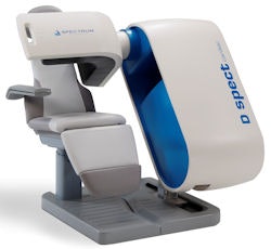
The D-SPECT camera uses nine collimated detector columns arranged in a curved configuration to conform to the shape of the left side of a patient's chest. The tungsten collimators are shorter (21.7 mm) and have larger square holes (2.26 mm) than standard models (45-mm length and 1.6-mm holes) used in conventional SPECT. An array of cadmium zinc telluride (CZT) crystals is aligned behind each collimator column.
 |
| Spectrum Dynamics' D-SPECT allows the patient to sit at a 45° angle, while the detector housing remains still during the scan. Image courtesy of Spectrum Dynamics. |
"By choosing to spend more time directing the independent small detector heads of the D-SPECT camera toward regions of interest," the authors wrote, "one can allocate more time to collecting data from these regions at the expense of collecting fewer data from less important regions. This design is equivalent to having a single detector head, with each of its pixels having a different time for data collection."
Image reconstruction
Data acquired by the D-SPECT camera are reconstructed using an iterative algorithm based on the maximum-likelihood expectation maximization (MLEM) method. To accelerate the MLEM algorithm, the technology applies an ordered-subset expectation maximization (OSEM) approach.
"Maximum-likelihood strategies, in layman's terms, can be thought about as, 'What's the likely distribution of activity that produced the counts that I observed,' " Gambhir explained. "Why that is particularly important with this type of scanner is that you are not observing counts from all around the body. You are only observing counts from part of the body, but each detector is observing counts in a different way, sampling angles in the myocardial region."
The researchers performed several phantom studies comparing D-SPECT with conventional SPECT technology. In addition, 18 patients were scanned with both technologies.
Among the 18 patients were 15 men, 13 of whom had known coronary artery disease. The patients underwent one-day technetium-99m sestamibi stress-rest SPECT, followed by D-SPECT imaging within 30 minutes of the SPECT scan. Stress and rest acquisition times were 19 and 11 minutes, respectively, for SPECT and four and two minutes, respectively, for D-SPECT.
Imaging protocol
Imaging on the D-SPECT camera was performed with the patients sitting upright and arms resting to the side. Images on the conventional SPECT camera were acquired using a 180° elliptical orbit with the patients in a supine position and arms positioned over their heads.
Three independent nuclear medicine physicians with no affiliations to Spectrum Dynamics interpreted the images. In addition, four nuclear medicine physicians assessed the images for confidence of interpretation.
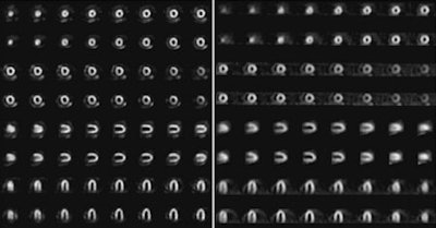
Overall image quality was rated good and higher in 17 (94%) cases for D-SPECT and 16 (89%) cases for SPECT. The study also noted that "D-SPECT images showed qualitatively better myocardial edge definition than did SPECT images in all cases, as assessed by four nuclear medicine physicians."
The three independent nuclear medicine physicians rated image quality as good and higher in 98% cases for D-SPECT and 92% cases for SPECT. D-SPECT images also showed qualitatively better myocardial edge definition than SPECT images in all cases.
 |
| The patient image compares D-SPECT (left) and conventional SPECT (right) tomographic slices in mid short axis (two levels), mid vertical long axis, and mid horizontal long axis. The top row of each set contains stress images and the bottom row shows rest images. Patient had no known cardiac history and had smoking as a risk factor; no perfusion defects are noted. Image courtesy of the Journal of Nuclear Medicine. |
D-SPECT's capabilities
Regarding the potential applications and capabilities of D-SPECT versus conventional SPECT, Gambhir said more human studies will have to be conducted, but "based on the improved spatial resolution, you would pick up smaller defects [with D-SPECT] that would be missed on a regular SPECT camera. You possibly could pick up more subtle changes between rest and stress."
Based on the limited number of patients in this study, he added that the results using D-SPECT are "more about seeing the same things as a regular SPECT camera, but in one-tenth the time. In reality, since there is better spatial resolution, as you do more low defect imaging studies, where someone has a mild stress-induced ischemia, you will pick that up better on the D-SPECT."
Business plan
Spectrum Dynamics' received 510(k) clearance from the U.S. Food and Drug Administration (FDA) for the D-SPECT in late 2006. More recently, the D-SPECT received the CE Mark in Europe and regulatory clearance for sales and marketing in Canada.
Over the past 18 months, the company has focused on completing a larger multicenter trial, with results expected to be published this year, and began installations of commercial systems in early 2008. Currently, there are 22 installations in the U.S. and one in Europe.
Spectrum Dynamics also recently received additional significant financing to market D-SPECT in Europe and to advance product development into noncardiac applications. Over the next two years, the company will expand the technology into oncology and other noncardiac applications.
"Rather than a technology change, the company expects a new design for the gantry and possibly larger detectors to cover a larger surface area for oncology applications," said Josh Gurewitz, Spectrum Dynamics vice president of sales and marketing. "It may be a whole-body camera or just a larger field-of-view, but that has not been determined."
By Wayne Forrest
AuntMinnie.com staff writer
April 10, 2009
Related Reading
Road to RSNA, Molecular Imaging, Spectrum Dynamics, October 24, 2007
Spectrum Dynamics completes Vanderbilt installation, April 25, 2007
Spectrum Dynamics receives 510(k) for D-SPECT, November 27, 2006
Road to RSNA, Molecular Imaging, Spectrum Dynamics, November 15, 2006
Spectrum Dynamics prepares launch of D-SPECT cardiac gamma camera, June 19, 2006
Copyright © 2009 AuntMinnie.com


