Researchers from Harvard Medical School in Boston used FDG-PET in mice to monitor a potential new treatment for Peutz-Jeghers syndrome, a rare, inherited disease that leaves patients predisposed to colon cancer and polyps for which they often undergo repeated surgeries.
Peutz-Jeghers syndrome results from a mutation in the tumor suppressor LKB1. People who inherit the flawed LKB1 gene have a 15-fold greater risk of developing colorectal cancer compared to the general population, and the mutation is a factor in many cervical carcinomas and non-small cell lung cancers as well (Proceedings of the National Academy of Sciences online, June 15-19, 2009).
Study authors David Shackelford, Ph.D.; Reuben Shaw, Ph.D.; and colleagues found that glucose avidity of the precursor lesions in Peutz-Jeghers patients revealed their existence on FDG-PET imaging before they had progressed to colorectal cancer.
FDG-PET imaging enabled the group to monitor lesion growth in mice during treatment with rapamycin analogs, a class of drugs that could someday be used to suppress the growth of lesions in Peutz-Jeghers patients.
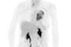
FDG-PET imaging was used on a microPET scanner to scan for the presence of polyps on the mutant mice. The polyps were found to have a high FDG avidity, and autopsy confirmed that polyps had caused the signal.
"Although most often used in the detection of malignant tumors, our findings suggest that FDG-PET may find clinical utility for the identification of polyps in [Peutz-Jeghers syndrome] patients," Shackelford and colleagues concluded.
FDG-PET could potentially be useful in determining whether secondary cancers that arise in Peutz-Jeghers patients at other sites (breast, pancreas, and endometrium) can also be visualized by FDG-PET, according to the authors.
Related Reading
Ultrasound screens for pancreatic cancer in high-risk populations, January 19, 2009
Capsule endoscopy detects small polyps in small intestine, May 16, 2005
Copyright © 2009 AuntMinnie.com


