Researchers from the University of California, Davis (UCD) have developed a dedicated breast PET/CT scanner they believe could be an improvement over breast imaging with whole-body PET or positron emission mammography. They describe their preliminary results with the system in the September issue of the Journal of Nuclear Medicine.
The new system combines work that had been conducted independently on both breast CT and breast PET at UCD Medical Center in Sacramento, CA. In addition to producing 3D images without having to compress a patient's breast, the technology could also be used as an extremity scanner for patients with rheumatoid arthritis (JNM, September 2009, Vol. 50:9, pp. 1401-1408).
"Although we list it as a dedicated breast scanner, we are billing it more now as an extremity scanner," said lead author Spencer Bowen, a graduate student in UCD's biomedical engineering program.
"The device is fully three-dimensional, so you can browse through the 3D structure of the breast and you don't have this shadowing problem [of 2D images]," added co-author and co-developer Ramsey Badawi, Ph.D., associate professor of radiology at UCD. "For example, that can be very problematic with mammography in patients with breast implants, because the implant will cover the cancer and make it difficult to visualize."
Homegrown technology
Both the CT and PET portions of the system were developed at UCD. Study co-author Simon Cherry, Ph.D., a professor in the biomedical engineering department, and his group began developing a dedicated breast PET scanner about 10 years ago. Co-author John Boone, Ph.D., a professor in the radiology department, has led a separate group on the development of a dedicated breast CT scanner for the past eight years.
About four years ago, Badawi and Bowen assembled their team to combine the two UCD-created imaging technologies. All the software, interfacing, gantry mechanisms, PET detectors, and associated electronics also were created at UCD.
The PET/CT system features a lutetium oxyorthosilicate-based dual-head PET camera and 768-slice conebeam CT scanner with a cesium iodide flat-panel detector (PaxScan 4030CB, Varian Medical Systems, Palo Alto, CA), tungsten target x-ray tube (Comet, Flamatt, Switzerland), and custom-made gantry that rotates 360° to acquire a full image.
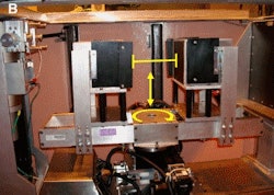 |
| UCD's PET gantry allows for control of detector height (vertical arrow), separation distance (horizontal line), and rotation (circular arrow). Images courtesy of UCD and the Journal of Nuclear Medicine. |
The PET and CT portions of the system also can be used independently.
Two years ago, the researchers initiated a clinical trial with women at high suspicion of having breast cancer as determined through mammography. Currently, a total of four patients and seven breasts (one patient had undergone a prior mastectomy) are included in the study.
Eligible patients were between 35 and 80 years of age, did not have a recent breast biopsy, and were not pregnant or diabetic. Before an injection of FDG, ranging from 174 to 477 MBq, patients fasted for more than four hours and were checked to ensure normal blood glucose levels of 200 mg/dL.
A CT scan was obtained, with the patient coached to perform breath-holds. A technician also used a hand controller to position the PET heads as close as possible to the patient's chest wall. The patient was advised to breathe normally and was scanned for 12.5 minutes by PET, with an average uptake time of 81 minutes.
"This technology takes us into a realm we could not go before," Badawi said. "Mammography screening turns up many patients with primary breast cancers that are 1 cm across or less, and this is too small for whole-body PET/CT. We expect the UCD breast PET/CT to pick up lesions half that size."
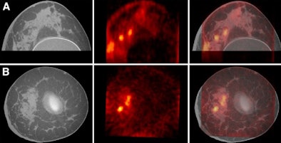 |
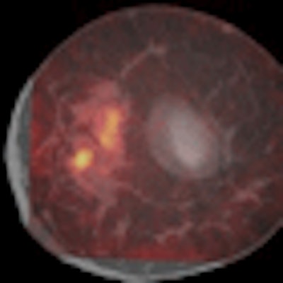
| Axial (A) and coronal (B) images from the UCD PET/CT system show CT (left), PET (middle), and fused breast images (right). |
Badawi believes the device could prompt a new paradigm for both breast cancer and rheumatoid arthritis treatment management.
Today, most patients with breast cancer are biopsied and then receive adjuvant chemotherapy. "Chemotherapy cuts the recurrence rate substantially, but many patients would not have recurred anyway," he said. With the dedicated PET/CT system, the patients potentially could be treated with a range of drugs before a tumor is removed to see if it responds and to determine which drug will be most effective. Similarly, by imaging the wrist, it may be possible to rapidly determine the effectiveness of new treatments for rheumatoid arthritis.
"One of the advantages of using PET versus mammography or any other anatomic imaging modality is that you can chart these changes much more rapidly with PET," Bowen added.
Future generations
With funds from the Susan G. Komen Breast Cancer Foundation, the researchers are working on a second prototype with enhanced detectors. The group also has funding from the U.S. National Cancer Institute (NCI) to build a third prototype, "which will have substantially improved detectors," Badawi said.
"We expect the next generation will have as big a difference, in terms of image quality, to the current generation as the current [PET/CT scanner] has to whole-body scanners," he said. "It should be a very significant step up."
Boone and his colleagues also have NCI funding to build a new-generation CT scanner, which will be combined with the next-generation PET camera. The new CT scanner will have slip-ring technology capable of dynamic CT, as well as higher resolution.
UCD plans to initiate two trials in the next six months to see if the technology can adequately chart therapy in breast cancer.
Eventually, the researchers plan to commercialize the system. "Whether we do that ourselves or have a company do that with us is not decided at this stage," Badawi said.
By Wayne Forrest
AuntMinnie.com staff writer
September 1, 2009
Related Reading
FDG-PET/CT predicts radiation-induced lung toxicity, July 1, 2009
PET/MRI breast imaging prototype shows early promise, June 16, 2009
FDG-PET/CT helps track pediatric bone and soft-tissue tumors, June 16, 2009
PET/CT shows early chemo results, April 16, 2009
FDG-PET/CT helps predict osteosarcoma survival, March 12, 2009
Copyright © 2009 AuntMinnie.com


