Friday, December 4 | 10:40 a.m.-10:50 a.m. | SST05-02 | Room E353C
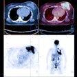
Call it a clash of imaging titans: 3-tesla MRI and PET/CT face off over the staging of colon cancer patients in this study to be presented in the scientific sessions by Italian researchers, who found that PET/CT is more accurate than whole-body MRI.The analysis covered 40 consecutive patients with previously diagnosed colon cancer. The patients underwent 3-tesla whole-body MRI (Achieva, Philips Healthcare, Andover, MA) and scanning with a whole-body FDG-PET/CT system (Discovery ST, GE Healthcare, Chalfont St. Giles, U.K.) for staging of lymph node and distant metastases after resection of the primary tumor.
Whole-body MRI correctly determined regional lymph node involvement in 75% of the cases, while PET/CT detected lymph node involvement in 90% of the patients. Whole-body MRI correctly staged distant metastases in 85% of the patients, compared with 90% for PET/CT.
"PET/CT reveals more hepatic metastases in colorectal cancer by identifying lesions as small as 4 mm," said lead author Dr. Guglielmo Manenti in the department of diagnostic imaging and interventional radiology at the University of Rome. "There always is a good correlation between morphologic imaging of whole-body MRI and functional imaging of PET/CT regarding evaluation of intralesional necrosis and peripheral activity."
Whole-body MRI, meanwhile, shows superiority in detecting metastasis in liver, brain, and bone, the researchers found.


