Researchers at Washington University School of Medicine in St. Louis are using PET scans on mice to test the efficacy of drugs to increase levels of dopamine in the brain and possibly alleviate some forms of attention deficit disorder (ADD).
The study, published online in Experimental Neurology, builds upon research from a year ago that found that higher dopamine levels in mice alleviate attention deficits caused by neurofibromatosis type 1 (NF1). Approximately 60% to 80% of children with NF1 have some type of attention deficit problem.
In the new study, co-author Dr. David Gutmann, PhD, director of the Washington University Neurofibromatosis Center, collaborated with Robert Mach, PhD, professor of radiology, on the use of the imaging agent raclopride, which binds to dopamine receptors in the brain and can be detected by PET.
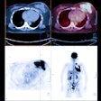
When raclopride was used to test dopamine levels in untreated mice, lower levels of brain dopamine allowed for greater raclopride binding, creating a brighter PET image. Following Ritalin treatment, the raclopride binding decreased.
The finding suggests that raclopride PET imaging could be used for preclinical testing of drugs that may affect brain dopamine levels. In addition, images can be obtained in an hour, enabling researchers to assess the effects of the drug on mouse behavior in a day.
Gutmann said the mouse model may not be perfect for studying all forms of attention deficit, but it is a terrific model for this type of attention system dysfunction. "Greater understanding of what goes wrong in some children with NF1 could lead to new insights into a broader variety of attention problems," he added.


