The combination of PET myocardial perfusion imaging and quantification of coronary flow reserve (CFR) can detect coronary artery disease (CAD), according to research presented at the Society of Nuclear Medicine (SNM) annual meeting in Miami Beach, FL.
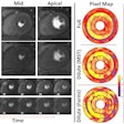
Swiss researchers found that quantifying CFR via PET myocardial perfusion imaging provided a significant advantage in unmasking CAD, even in patients who would otherwise be considered healthy with normal myocardial perfusion imaging.
In a study involving 73 cases, a team led by Dr. Philipp Kaufmann of University Hospital Zurich found that myocardial perfusion imaging with PET and either rubidium-82 or nitrogen-13 ammonia with quantitative CFR measurements significantly improved the sensitivity and diagnostic value of stress testing over myocardial perfusion imaging alone.
In a separate study from Brigham and Women's Hospital and Harvard Medical School involving 704 patients older than 75, researchers used similar methods and concluded that age was not necessarily a risk factor for developing CAD.
Lead investigator Dr. Venkatesh Murthy, PhD, said the research showed that many older adults have preserved coronary vascular function and the group has an extremely favorable prognosis. The results suggest that the loss of vascular function may not be an inevitable consequence of aging, Murthy said.


