For NYU Langone Medical Center in Manhattan, the acquisition of a new PET/MRI scanner was a no-brainer. From a clinical perspective, the hybrid modality's ability to simultaneously image metabolic and physiological functions provides a platform for the facility to expand its research capabilities and offer better care for patients.
"In my view, it is a story of convergence -- a convergence of imaging modalities and a convergence of disciplines, specialties, and agendas," said Dr. Daniel Sodickson, PhD, vice chairman of research in NYU Langone's departments of radiology, physiology, and neuroscience. "It will be a large part of our expansion from the MR side into the PET side. If you put those two together, you have anatomic, microstructural, functional, and metabolic information all in one modality."
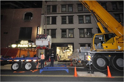
NYU Langone installed its PET/MRI scanner (Biograph mMR, Siemens Healthcare) in May at its Bernard and Irene Schwartz Center for Biomedical Imaging, of which Sodickson is the director. The system is next to a 7-tesla MRI scanner and close to two 3-tesla MRI scanners.
 |
| Siemens' Biograph mMR PET/MRI arrives at NYU Langone. All images courtesy of NYU Langone Medical Center. |
Structural modifications
The room in which the PET/MRI system is located previously held an MRI scanner; NYU Langone renovated the entire room to accommodate PET functions for the device. Modifications were made for radiotracer handling, including injection of the radiotracers, a waiting area for patients who receive the radiotracers, and a "hot" restroom for patients.
"This is standard practice for PET and PET/CT already, but we set this area aside to make sure all the other staff working in the area and patients do not get any unnecessary exposure to radioactivity," Sodickson said. "Obviously, the doses that the patients are getting are still safe, but they are merited by the potential benefit of the scan."
NYU Langone declined to disclose the monetary investment in the renovations, but the cost "was not incredibly expensive," Sodickson said. "The modifications so far are minor. The more significant modifications will occur when we have our cyclotron and our radiochemistry laboratory."
Clinical applications
Initially, NYU Langone plans to target neurologic, oncologic, and musculoskeletal applications for its new PET/MRI service.
Neuroimaging has been an area of great attention for both PET and MRI, taken separately. PET, combined with new, novel radiotracers, is showing promise in advancing the understanding of neurodegenerative disease, such as Alzheimer's and multiple sclerosis, and various psychiatric afflictions. In cancer cases, both MRI and PET have been used extensively to identify and characterize tumors.
One great interest within musculoskeletal studies is tracking inflammation. "There are some preliminary PET markers that may combine with MRI markers, so we can use that information in a complementary way, but that is more of a 'blue sky' project for the future," Sodickson said. "Perhaps we can start making a dent in musculoskeletal disease, which is a huge public health problem."
 |
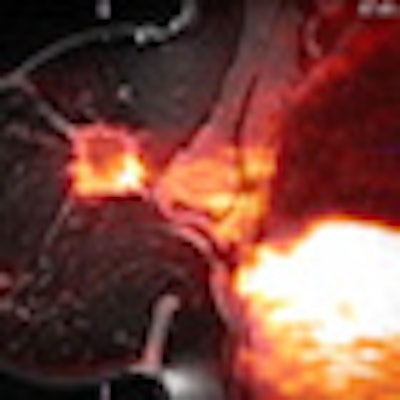
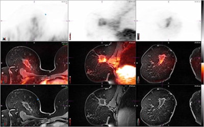
| Localized breast image of a female patient in her 50s. FGD-PET image shows increased FDG uptake, which could represent residual malignancy versus postsurgical change. Fusion with the anatomically detailed T1-weighted postcontrast MR image shows increased uptake localized to a smoothly marginated seroma and is benign. |
First PET/MRI scan
NYU Langone scanned its first patient on the PET/MRI system in late July. The protocol called for the patient to undergo a PET/MRI exam after a PET/CT study to compare the performance of PET/MRI with PET/CT in detecting whole-body disease. In this case, the scans were performed for metastatic breast cancer.
The procedure is part of a larger study in which the facility is enrolling breast cancer patients who are undergoing FDG-PET/CT to assess systemic disease, either at the time of diagnosis or during or after therapy. The goal is to determine if PET/MRI can provide additional information to detect brain and liver metastases. One benefit is that PET/MRI exposes a patient to one-third the radiation dose of PET/CT. The principal investigator is Dr. Amy Melsaether, an assistant professor in the department of radiology.
"In the case of breast cancer, there are two methods by which lesions are usually characterized -- one with [MRI] diffusion imaging, which tracks the number of cells and the density of cells and is one measure of malignancy," explained Eric Sigmund, PhD, an assistant professor of radiology. "On the PET side, the radiotracer FLT [fluoro-L-thymidine] is sensitive to the growth rate of cells. Those are malignant markers, but they are not equivalent, although they are treated that way clinically. The ability to measure both of them simultaneously [with PET/MRI] will be very powerful and allow us to classify lesions in ways we have not been able to before."
Enthusiastic welcome
Even before the PET/MRI system arrived, there was "a remarkable groundswell of interdisciplinary collaboration and interest," Sodickson said. "We got concrete buy-in from 10 clinical departments, each of whom said, 'Not only are we interested in this, but our clinicians will want to send patients and our investigators will want to work on it. We want to chip in to support this program as an institutional resource.' This generally does not happen when a piece of apparatus arrives at a medical center. This was the snowball going downhill."
To that end, NYU Langone created an interdepartmental steering committee to accelerate the integration of the PET/MRI system into clinical practice. The hybrid imager has also changed the way NYU Langone recruits physicians both inside and outside of the facility.
"It used to be that everyone would recruit individually," said Dr. Michael Recht, chairman of the department of radiology. "Now when we recruit somebody, the question is whether they help collaboratively across the institution and across the program. We have been looking at some of our hematology and oncology recruits to see how they fit into our program, and they are looking into some of our recruits."
Physicians outside of NYU Langone have also expressed an interest in joining the facility, largely because of the new PET/MRI scanner, which is the first such hybrid device in the New York City area. "The key, of course, will be to come up with the diagnostic and therapeutic techniques to draw in the patients who need it," Sodickson added.
Logistical planning
NYU Langone has held weekly meetings with a planning group to tackle a number of PET/MRI-related issues, including appropriate training, scheduling scans, and patient logistics.
There has been general training on how staff should handle themselves around radioactive PET tracers, which is "mostly stay away from them unless you are one of the authorized users," Sodickson said. "We have also devoted a lot of thought to which technologists will be running the scans."
That initiative has prompted nuclear medicine technologists and MRI technologists to combine and share their respective expertise. Once NYU Langone starts to get more hybrid images, the facility plans to expand basic research and training in the interpretation of the PET/MRI images.
PET/MRI reimbursement
On the issue of PET/MRI reimbursement, NYU Langone has been working with insurance companies to develop an appropriate model for when the imaging exams are performed simultaneously.
Recht's understanding is that the U.S. Centers for Medicare and Medicaid Services (CMS) has said that if a patient receives a PET/MRI scan, the PET portion of the scan can be reimbursed under the PET code. If the same patient undergoes a medically necessary MRI exam, a facility can bill for the portion of the MRI scan that is medically appropriate.
"My understanding is that there has not been a single patient who has been reimbursed for the [PET/MRI] study yet, so I think it is still a work in progress," Recht added. "Ideally, we will be able to get CMS to create CPT codes for the hybrid study, which is appropriate, as [CMS] has done for PET/CT."


