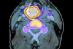
PET imaging with F-18 fluorothymidine (FLT) during treatment and early follow-up could predict therapeutic response and identify patients who need close follow-up to detect persistent or recurring head and neck squamous cell carcinomas (HNSCCs), according to a study published in the October issue of the Journal of Nuclear Medicine.
In the study from Kagawa University in Japan, 28 patients with HNSCCs underwent FLT-PET and FDG-PET imaging prior to treatment with radiation therapy, four weeks after the start of therapy, and five weeks after the conclusion of therapy. Uptake of both of the agents was measured in primary and metastatic lesions.
During the radiation therapy, FLT uptake disappeared in 34 (63%) of 54 lesions, with a negative predictive value of 97%. FDG uptake also had a high negative predictive value of 100% during radiation therapy, but only nine lesions (16%) showed absence of FDG.
In addition, the specificity and overall accuracy of FLT-PET were significantly higher than those of FDG-PET during and after radiation therapy.
FLT-PET is more useful for assessing early locoregional clinical outcomes and is helpful for avoiding unnecessary radical surgery, lead study author Dr. Hiroshi Hoshikawa and colleagues concluded.


