The radiotracer carbon-11 (C-11) methionine (MET) with PET/CT can help clarify negative or inconclusive FDG-PET findings in multiple myeloma patients, as well as differentiate between atypical and typical pituitary adenomas, according to two Japanese studies presented at last week's RSNA 2012 conference.
In one study, researchers from Southern Tohoku General Hospital performed both FDG-PET/CT and MET-PET/CT scans on 69 patients with pituitary adenomas. The patient group consisted of 17 men and 52 women with a mean age of 42.8 years (range, 26 to 59.6 years).
MET-PET was performed 20 minutes after injection of the tracer, said lead study author and presenter Dr. Noriaki Tomura. FDG then was injected 60 minutes after MET-PET imaging, with the FDG-PET exam performed 60 minutes after FDG injection. Images were obtained at five or 10 minutes per table position.
Tomura and colleagues measured maximum standardized uptake value (SUVmax) for each adenoma. SUVmax of the pituitary gland also was calculated for 42 patients with suspected brain tumors, who underwent both FDG-PET/CT and MET-PET/CT and acted as controls with normal pituitary glands. Their tumors were not located near the pituitary gland, hypothalamic region, or optic chiasm, the researchers noted.
Mean SUVmax with MET and FDG differed significantly between normal glands and typical adenomas and between typical and atypical adenomas, Tomura and colleagues found. The mean SUVmax with MET was 2.1 (± 0.9) in normal glands and 3.5 (± 2.2) in typical adenomas. By comparison, mean SUVmax with FDG was 3.3 (± 0.7) in normal glands and 5.5 (± 6.4) in typical adenomas.
In atypical adenomas, the mean SUVmax with MET was 5.9 (± 2.4), compared with a mean SUVmax of 13.5 (± 12.1) with FDG.
Based on the results, Tomura and colleagues concluded that both FDG-PET/CT and MET-PET/CT can provide useful information in the diagnosis of pituitary adenomas and can help determine the possibility of atypical adenoma before surgery.
Multiple myeloma
In the second study, lead author Dr. Yuji Nakamoto, PhD, and colleagues from Kyoto Graduate School of Medicine evaluated 20 patients with histologically proven multiple myeloma. The 11 men and nine women (average age, 61 years) underwent a total of 16 FDG-PET/CT and MET-PET/CT scans within three weeks of each other for either staging or restaging of the disease.
Whole-body PET/CT was performed 20 to 30 minutes after injection of MET and one hour after administration of FDG. Both sets of images were analyzed visually on a scale of 0 to 4, with 0 as negative and 4 as positive.
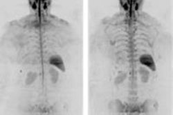
Nakamoto and colleagues confirmed active myeloma in 11 of the 16 studies, including two patients with extramedullary lesions. Consistent results were obtained in 12 studies, with six positive and six negative results. In five studies of grade 4 for both MET and FDG scans, MET-PET discovered more lesions than FDG-PET.
MET-PET was positive in all six studies with hypergammaglobulinemia, Nakamoto noted; however, abnormal MET uptake was seen in four of 10 patients who did not have hypergammaglobulinemia.
Excluding two patients whose final diagnoses could not be determined, the patient-based analysis found that sensitivity for MET was 91%, compared with 82% for FDG. MET also achieved greater accuracy of 93%, compared with 86% for FDG. MET and FDG both reached specificity of 100%.
"MET-PET/CT revealed comparable or more lesions clearly, as compared with FDG-PET/CT in multiple myeloma," the researchers concluded. "It could be helpful especially when negative or inconclusive findings are obtained on FDG in spite of highly suspicious of recurrent myeloma."


