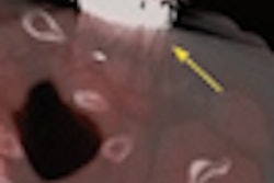
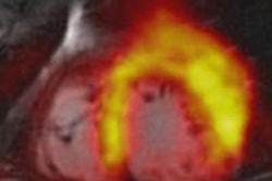
Researchers from Mount Sinai Hospital are testing a new sugar-based radiotracer known as fluorodeoxymannose (FDM) as a cardiovascular PET imaging agent.
The study, published online January 12 in Nature Medicine, compared FDM with FDG for imaging inflamed high-risk vulnerable artery plaques before they rupture.
Principal investigator Dr. Jagat Narula, PhD, director of Mount Sinai's cardiovascular imaging program, said preclinical tests show that PET imaging with FDM may offer a more targeted tool to detect dangerous plaques and inflammation that may be associated with serious cardiovascular events.
While early results indicate that FDM performs comparably to FDG, the radiotracer may be able to more specifically target inflammation due to plaque-infiltrating macrophages that develop mannose receptors (MRs), according to Narula.
The results show that mannose is taken up by this specific subset of macrophage cells that dwell in high-risk plaques.
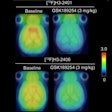
In the study, FDG and FDM were compared using PET in atherosclerotic animal models. While uptake of each tracer within atherosclerotic plaques and macrophage cells was similar, the experimental FDM tracer showed at least 25% greater FDM uptake due to MR-bearing macrophages.


