Retinal imaging technology may provide highly predictive early detection of changes associated with Alzheimer's disease, according to research being presented July 15 at the Alzheimer's Association International Conference in Copenhagen, Denmark.
Preliminary findings from an Australian clinical trial show that the optical imaging technology, which was created at Cedars-Sinai Medical Center and licensed to start-up NeuroVision Imaging, can detect changes that occur 15 to 20 years before clinical diagnosis of Alzheimer's.
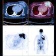
PET scans use radiotracers, and cerebrospinal fluid analysis requires that patients undergo invasive and often painful lumbar punctures; neither approach is feasible, especially for patients in the earlier stages of disease, study co-author Dr. Keith Black, professor and chair of Cedars-Sinai's department of neurosurgery, said in a statement.
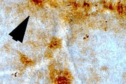
Beta-amyloid plaques associated with Alzheimer's disease occur in the brain and also in the retina, Black noted. By "staining" the plaque with curcumin, a component of the common spice turmeric, it can be detected in the retina before it begins to accumulate in the brain. The optical imaging device allows the researchers to look through the eye, as an ophthalmologist would, to see these changes.
The preliminary results from 40 patients showed the technology could differentiate between Alzheimer's disease and non-Alzheimer's disease with 100% sensitivity and 81% specificity.


