A new technique for imaging and measuring how quickly blood flows in the brain could help researchers better understand and treat drug abuse, according to a study published in Biomedical Optics Express.
Researchers from Stony Brook University and the U.S. National Institutes of Health used optical coherence Doppler tomography (ODT) to measure how cocaine disrupts blood flow in the brains of mice. Lead author Jiang You and colleagues were able to identify cocaine-induced microischemia -- a precursor to stroke (Biomed Opt Express, September 1, 2014, Vol. 5:9, pp. 3217-3230).
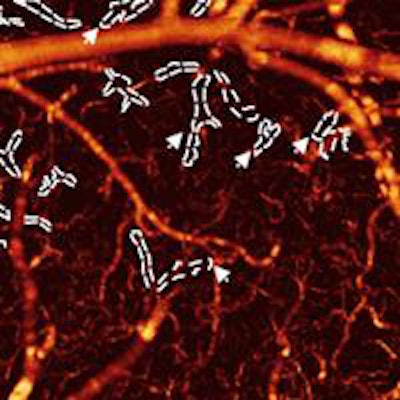
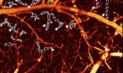
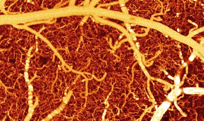 The images show blood flow in a healthy mouse brain versus a mouse brain exposed to cocaine. The image above shows the mouse brain blood vessels before cocaine. The image below shows the blood vessels after cocaine; many of the vessels are now darker, signifying lower blood flow. Field-of-view: 1.2 x 2.0 x 1.0 mm3. Images courtesy of Biomedical Optics Express.
Techniques such as functional MRI (fMRI) provide a good overall map of the flow of deoxygenated blood, but they do not have high enough resolution to study what happens inside the capillaries, according to the authors. ODT works with small blood vessels; with the technique, laser light hits and bounces back from moving blood cells. Researchers then measure the shift in the reflected light's frequency to determine how fast the blood is flowing.


