Sunday, November 30 | 12:05 p.m.-12:15 p.m. | SSA13-09 | Room E451B
German researchers are lauding the ability of PET/MRI to provide "superior assessment" of therapy based on functional metabolic response, but morphologic information falls short in evaluating therapeutic response in soft-tissue sarcoma patients after isolated limb perfusion.Given that PET/MR images and morphologic, molecular, and metabolic information are obtained simultaneously, researchers believe that a single scan may be sufficient to grade a tumor, determine tumor blood flow, and gauge therapy response.
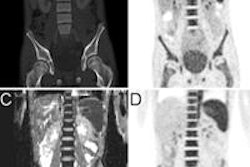
As a result, researchers from University Hospital Essen evaluated 10 patients with histologically proven soft-tissue sarcomas who underwent FDG-PET/MRI scans (Biograph mMR, Siemens Healthcare) prior to and six weeks after isolated limb perfusion.
All tumor lesions were assessed from the baseline and follow-up exams to determine maximum standardized uptake values (SUVmax) for metabolic results and morphologic response through the maximum diameter according to the Response Evaluation Criteria in Solid Tumors (RECIST).
"The potential clinical benefits for patients are that adding the metabolic response criteria may lead to more accurate therapy monitoring, which is very important for further clinical workflow," lead author Dr. Michael Herbrik said.
Three patients responded to treatment, while seven did not respond, the researchers found. The three responders exhibited a significant decline in SUVmax, while the nonresponders had considerably less change in SUVmax.
While PET/MRI enables "superior assessment" of therapy based on a patient's functional metabolic response, morphologic assessment using RECIST evaluation does not provide "sufficient evaluation of therapy response in soft-tissue sarcomas cases with isolated limb perfusion," the group concluded.
Herbrik and colleagues plan to continue their research to gather more reliable data for specific subforms of sarcoma.


