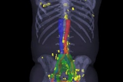
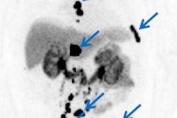
PET performed with a new imaging agent finds more prostate cancer than other modalities, including CT, MRI, and PET with conventional radiotracers, according to a study presented at this week's Society of Nuclear Medicine and Molecular Imaging (SNMMI) meeting.
The new radiopharmaceutical detects prostate cancer that has spread to other tissues by zeroing in on prostate-specific membrane antigen (PSMA), which is associated with prostate cancer. The radiotracer enables clinicians to detect prostate cancer cells that have metastasized to bone, said the researchers from Memorial Sloan-Kettering Cancer Center.
The new radiotracer combines small amounts of zirconium-89 (Zr-89) with a piece of an antibody known as a minibody, which has anti-PSMA qualities and attaches to overexpression of the enzyme. The radiotracer, known as Zr-89 Df-IAB2M (IAB2M), is imaged faster with PET than the full antibody (J591) that targets PSMA.
In the study, 28 subjects were imaged with CT, MRI, and bone scintigraphy, as well as conventional FDG-PET. PET with the experimental Zr-89 agent was evaluated in escalated doses.
The researchers found 393 suspected lesions in soft tissue using all imaging modalities together. PET/CT with Zr-89 identified 81.7% of all suspected bone lesions, including 65 that were not identified by any other modality, according to the study team. The radiotracer also found 32 soft-tissue lesions not detected by other methods.
Initial results of full-body imaging with Zr-89 demonstrate that the new agent detects more disease sites than conventional imaging, the group concluded. Further validation could enable the radiotracer to be used for specific targeted biopsies, leading to earlier and better treatment for prostate cancer patients.


