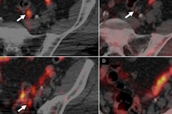
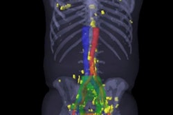
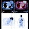
PET/CT scans with a radiotracer that targets prostate-specific membrane antigen (PSMA) are significantly more effective than other detection methods currently in use.
That conclusion comes from a study published in the January issue of the Journal of Nuclear Medicine, in which researchers from Johns Hopkins Medical Institutions compared PET/CT with F-18 DCFBC to conventional imaging modalities, such as technetium-99m methylene diphosphonate (MDP) bone scans and contrast-enhanced CT of the chest, abdomen, and pelvis.
The lesion-by-lesion analysis of 17 patients found that DCFBC-PET detected 592 positive lesions, compared with 520 positive lesions with the conventional methods (JNM, January 2016, Vol. 57:1, pp. 46-53).
DCFBC-PET also achieved greater sensitivity (92%) for detecting prostate cancer lesions in lymph nodes, bone, and visceral tissue than current methods (71%).


