A work-in-progress hybrid optical-gamma imaging system can provide a visual representation of molecular data in the same frame as optical images of surface anatomy such as the skin and eyes, according to research presented at this week's Society of Nuclear Medicine and Molecular Imaging (SNMMI) conference in San Diego.
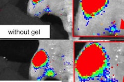
A team from the University of Nottingham in the U.K. has paired a gamma camera with an optical imaging system to create a portable hybrid molecular imaging device. In a clinical pilot study, the handheld hybrid system effectively imaged lymphatic and thyroid tissue as well as drainage from the lacrimal glands, according to the group led by Alan Perkins, PhD.
"Successful absorption of the radionuclides in these targeted areas was clearly seen in tandem with optical images of surface anatomy," Perkins said in a statement from the SNMMI.
The device includes a 1.5-mm-thick scintillator and a 1-mm pinhole collimator, which allowed the investigators to optimize image resolution and keep acquisition time under five minutes, according to the society. In their pilot study, the investigators imaged subjects who were undergoing routine molecular imaging procedures such as bone scans or imaging of the thyroid, eye, or lymphatic system.
"This scanner has handheld potential and can be used in a variety of settings, including the outpatient clinic, patient bedside, operating theatre, and intensive care unit," Perkins said.
The researchers noted that the system is still being developed and will require further testing before being made available to wider patient populations.


