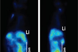
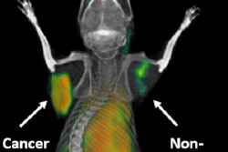
Researchers at Stanford University are assessing how well new PET radiopharmaceuticals, called immuno-PET tracers, can accurately identify molecules in cancer cells that prevent the immune system from attacking the disease, according to a study published in the April issue of the Journal of Nuclear Medicine.
While the promising preliminary results come from animal models, the researchers believe their efforts ultimately will be effective for human imaging, with immuno-PET tracers able to help patients receive optimal treatment (JNM, April 2017, Vol. 58:4, pp. 538-564).
Immune checkpoint inhibitors have emerged as a promising approach for treating cancer. However, the lack of imaging methods to assess immune checkpoint expression has been a major stumbling block to predicting and monitoring response to a clinical checkpoint blockade.
In the study, the researchers examined immuno-PET radiotracer design modifications and their effects on human immune checkpoint imaging. By optimizing design parameters, they hope to develop a noninvasive molecular imaging tool for eventual monitoring of clinical checkpoint blockade.
"Using animal models, this study shows the development of several new engineered PET tracers that can help image the immune system in action and be used to monitor checkpoint inhibitor therapy," senior author Dr. Sanjiv "Sam" Gambhir, PhD, said in a statement.


