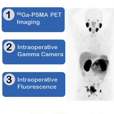
DENVER - The Society of Nuclear Medicine and Molecular Imaging (SNMMI) on Wednesday awarded its Image of the Year to researchers who used PET/CT followed by fluorescence-guided surgery to demonstrate how dual-labeled prostate-specific membrane antigen (PSMA) inhibitors based on PSMA-11 enhance preoperative staging of metastatic prostate cancer.
The combined imaging approach, by researchers from the German Cancer Research Center (DKFZ) and Heidelberg University Hospital, is designed to produce more accurate detection of PSMA-positive tumor lesions and improve the removal of lymph-node metastases using image-guided surgery, leading to better patient outcomes.
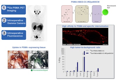
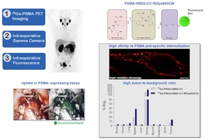
Images show dual-labeled PSMA inhibitors for prostate cancer with PET/CT and fluorescence-guided intraoperative identification of metastases. The technique might establish a new treatment regimen for more precise and sensitive detection before, during, and after therapy for prostate cancer. Image courtesy of the German Cancer Research Center and Heidelberg University Hospital.
"There has been a huge effort to improve care of prostate cancer patients using molecular imaging," said Dr. Satoshi Minoshima, PhD, chair of the SNMMI Scientific Program Committee and SNMMI vice president-elect. "The study presented clearly demonstrates that the combined PET imaging, gamma detection, and optical imaging can help not only preoperative staging of the disease but also intraoperative guidance of metastatic lymph-node dissection. We anticipate that such hybrid cancer detection methods will become prevalent in the near future and contribute significantly to the care and management of prostate cancer patients."
Lead author Dr. Ann-Christin Baranski from the German Cancer Research Center said she and her colleagues were "deeply honored to receive this award."
"I would like to thank all team members who contributed to this interdisciplinary work," she said. "As resection of lymph-node metastases has considerable impact on the outcome of metastatic prostate cancer patients, the aim of our study is to improve the intraoperative accuracy of detecting PSMA-positive tumor lesions."
The study was supported by the VIP+ funding program of the German Ministry of Education and Research.


