Researchers have developed a same-day, noninvasive PET imaging approach to assess tumors that have the programmed death ligand 1 (PD-L1) protein. The technique could potentially help guide cancer treatment decisions and evaluate treatment response, according to results published in the March issue of the Journal of Nuclear Medicine.
For the study, a team led by David Donnelly, PhD, of Bristol-Myers Squibb Research and Development labeled an adnectin protein that binds to PD-L1 with F-18. The group then used PET imaging to track the protein in mice with PD-L1 negative and PD-L1 positive subcutaneous tumors, as well as to evaluate its distribution, binding, and radiation dosimetry in a healthy monkey (JNM, March 1, 2018, Vol.59:3, pp. 529-535).
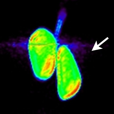
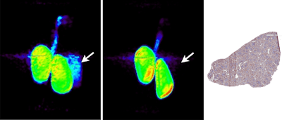
 Whole-body PET (maximum intensity projection) images of monkey kidneys and spleen 90 minutes postinjection of anti-PD-L1 adnectin labeled with F-18 (left) and of the labeled protein with co-administration of a nonlabeled dose in the same monkey (center). Right image shows the anti-PD-L1 immunohistochemistry of healthy monkey spleen tissue. Credit: David Donnelly, PhD.
The technique was feasible, the researchers found, and radiation dosimetry estimates showed that the tracer is safe to administer in human studies.
"This approach represents an opportunity for physicians to noninvasively assess all of a patient's tumors for PD-L1 expression with a single PET scan and timely readout," Donnelly said in a statement released by the journal. "This may help guide treatment decisions and assess treatment response, to help identify the right treatment for the right patient at the right time and right dose."
Clinical studies are now underway to measure PD-L1 expression in human tumors, the authors wrote.


