Augmented reality (AR) visualization of SPECT/CT data can help guide surgeons in identifying sentinel lymph nodes in patients with metastatic melanoma, according to a study published January 3 in Oral Oncology.
In a pilot study, a group of Japanese and U.S. researchers simulated sentinel lymph node metastases on head and neck Styrofoam phantoms. The team measured the time required for physicians to identify sentinel lymph node targets on the phantoms using a handheld gamma probe alone compared to the use of the gamma probe while wearing an AR headset.
The results suggest the combined method is accurate and that using the AR headset can significantly reduce intraoperative time, according to the authors.
"An augmented reality headset capable of displaying 3D virtual renderings of SPECT/CT allows localization of targets in the head and neck region within an average accuracy of 9 mm and may reduce the time needed to localize targets when paired with the conventional gamma probe method," wrote Dr. Ryusuke Nakamoto, deputy director of radiology at the Japanese Red Cross Wakayama Medical Center.
The pathologic status of sentinel lymph nodes is the most important prognostic factor for patients with clinical stage metastatic melanoma. Accurate guidance when removing the lymph nodes is essential for surgeons.
Preoperative SPECT/CT imaging allows surgeons to identify suspected cancerous lymph nodes in relation to other surrounding structures in 3D with high detail, while a handheld nonimaging gamma probe that detects uptake of the SPECT radiotracer is generally used in the operating room.
However, SPECT/CT imaging is viewed on an external computer monitor and surgeons must retain the visual data in memory as they look to and from the surgical field, the authors wrote.
In this study, the researchers tested an AR system they developed that projects a 3D virtual rendering of the SPECT/CT data onto patient phantoms using a wearable AR headset (HoloLens, Microsoft). They investigated whether the system could reduce detection time by combining the gamma probe with the AR system, as opposed to using the gamma probe alone.
The researchers painted eight head and neck Styrofoam phantoms with a mixture of technetium-99m (Tc-99m) radiotracer detectable with a handheld gamma probe and fluorescent ink visible only under ultraviolet (UV) light. They create between 10 and 20 simulated lymph nodes on the surface of the phantoms.
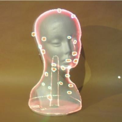
After obtaining SPECT/CT images of these phantoms, virtual renderings of the nodes were generated from the SPECT/CT data and displayed using the AR headset, which allows users to visualize the data directly in their line of sight.
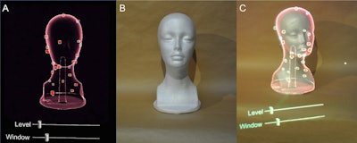
 Creating a 3D virtual rendering and superimposing it on the real world. After creating a 3D virtual rendering from SPECT/CT images (A), the 3D virtual rendering can be superimposed on a real-world mannequin (B) using voice commands on the AR headset. Bars were set to adjust the level and width of the window with hand gestures to optimize the visibility of simulated lymph nodes while wearing the AR headset (A, C). Image courtesy of Oral Oncology.
For each of three participating physicians, the researchers recorded the time required to localize lymph node targets using the gamma probe alone and using the gamma probe while wearing the AR headset. The surface localization accuracy when using the AR headset was evaluated by measuring the misalignment between the locations visually marked by the evaluators and the ground truth locations identified using UV stimulation of the ink at the site of the nodes.
For all three evaluators, using the AR headset significantly reduced the time needed to detect targets. The average time with the gamma probe alone was 30.5 seconds, compared with 11.2 seconds with the gamma probe and AR system combined. The combined use of the AR headset with the gamma probe significantly reduced the detection time for all three evaluators (p = 0.012), with an average percent time saved of 61% (range, 21%-86%).
In addition, the average misalignment between the location marked by the evaluators when using the AR headset and the ground truth location was 8.6 mm. Regions with the smallest and largest misregistrations were level III (7 mm) nodes and occipital nodes (12.7 mm).
"AR visualization of SPECT/CT data in 3D allows for accurate localization of targets in the head and neck region, and may reduce the localization time of targets," the authors wrote.
A combined SPECT/CT and AR approach could allow surgeons to directly visualize the position of the radioactivity emitted by sentinel lymph nodes without needing to translate SPECT/CT image information from a workstation outside of the surgical field of view to the patient, the researchers wrote.
Furthermore, even if several targets are in close proximity to each other, this 3D imaging technique has the potential to accurately distinguish each target, since it allows for multidirectional viewing, they added.
Ultimately, this study establishes initial data to guide future development of the method and enables planning for in vivo animal and clinical studies, the researchers wrote.
"More work is needed to improve the registration and AR display for use in clinical settings," the researchers concluded.


