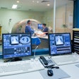
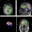
VIENNA - A group from the University of Rome in Italy used a polarized MRI phased-array coil in a new diagnostic approach for the biopsy localization of prostate nodules. Dr. Valeria Panebianco presented the results at the European Congress of Radiology on Tuesday
The diagnosis of prostate nodules can be divided into three stages, according to Panebianco. Prostate-specific antigens (PSA) are measured for screening, while detection is generally done with transrectal ultrasound (TRUS) or fiber-optic cystoscopy. Finally, biopsy and CT can be used for lesion characterization and staging.
"In this context, our question is that when we have a patient population with high PSA and doubtful or negative TRUS results, does MRI have a role?" Panebianco said.
In this study sample, 91 patients with high PSA values and negative, or doubtful, TRUS results were enrolled. MRI was performed with a 1.5-tesla Magnetom Vision Plus (Siemens Medical Solutions, Erlangen, Germany). Two-dimensional fast spin-echo axial, sagittal, or coronal images were acquired depending on the location of the lesion. A T1-weighted gradient-echo sequence also was used. The matrix of 420 x 512 was crucial for obtaining high-resolution images, Panebianco pointed out.
The total examination time was about 20 minutes. In terms of gland dimensions, seminal vesicle alteration and peripheral zone changes appeared as hypointense areas on T2-weighted images. The gland dimensions ranged from 40 to 70 mm. MR imaging also revealed neoplastic changes in 23 patients, such as the appearance of plaque, and inflammatory changes in bilateral strips in 40 patients, she said.
For staging, the group determined that 16.4% of the patients had capsular involvement, while 13.7% had seminal involvement. The remaining patients were diagnosed with lymphoadenopathy. The sensitivity of MR was 92%, the specificity was 66%, and the accuracy was 70.9%.
"The sites of cancer predicted by MRI were well-correlated with the sites of cancer at biopsy," Panebianco said. As a result, prostate imaging with MR has become a routine part of cancer diagnosis at her institution, she added.
By Shalmali Pal
AuntMinnie.com staff writer
March 6, 2001
Click here to post your comments about this story. Please include the headline of the article in your message.
Copyright © 2001 AuntMinnie.com


.fFmgij6Hin.png?auto=compress%2Cformat&fit=crop&h=100&q=70&w=100)


.fFmgij6Hin.png?auto=compress%2Cformat&fit=crop&h=167&q=70&w=250)


