In vivo proton MR spectroscopy of the breast continues to show encouraging results in its ability to link choline levels to carcinoma, as well as determine if the cancer has invaded the axillary lymph nodes, according to investigators in Hong Kong.
David Yeung, Ph.D., and colleagues from the Prince of Wales Hospital in Shatin, Hong Kong reported their findings in Radiology Online. Research from the past decade has associated high levels of choline-containing compounds (3.2 ppm) to malignant lesions on 1H MR spectroscopy.
The current study builds on Yeung’s previous research with MRS and breast lesions. The earlier work sought to assess the clinical usefulness of tagging choline with MRS. The results showed that choline could be reliably detected in less than 45 minutes in large contrast-enhanced breast lesions.
"Although the use of the presence of choline...has shown promising results, a number of false-negative findings involving various in-situ and invasive lesions also have been reported. It is unclear whether the failure to detect choline in some breast cancers is related to insufficient sensitivity or is an indication of less aggressive lesion types," they wrote (Radiology Online, August 2, 2002).
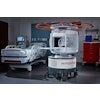
The latest study included 39 women with a mean age of 52.5 years who had histopathologically confirmed breast cancer. Imaging was performed on a 1.5-tesla whole-body MR system (Gyroscan ACS NT, Philips Medical Systems, Andover, MA).
Transverse images were obtained using a T1-weighted spin-echo sequence with spectral presaturation and inversion recovery for fat saturation. Then a bolus intravenous injection of gadopentetate dimeglumine (Magnevist, Schering, Berlin, Germany) was administered at 0.2 mmol per/kg, and contrast-enhanced images were acquired.
Unenhanced sagittal images of the affected breast and axillary lymph nodes were obtained with T2-weighted turbo spin-echo with spectral presaturation and inversion-recovery fat-saturation sequences. Contrast-enhanced sagittal images also were obtained.
Using point-resolved spectroscopic sequence (PRESS) with either a bilateral breast coil or a circular surface coil as the receiver, three water-suppressed spectra were acquired for each volume of interest.
"The signal-to-noise ratio of the apparent peak at 3.2 ppm was measured, and a minimal signal-to-noise ratio of 2 was chosen to indicate that choline was present in a spectrum," the authors explained.
All 39 breast lesions underwent core-needle biopsy; all axillary lymph nodes underwent ultrasound-guided fine-needle aspiration biopsy or lymph node dissection.
According to the results, 25 cancers were diagnosed as infiltrating ductal carcinoma (IDC); two as invasive lobular carcinoma; two as medullary carcinoma; two as mucinous carcinoma; three as IDC with an extensive in situ component; and four as ductal carcinoma in situ (DCIS). The median largest dimension of these contrast-enhanced breast lesions was 2.5 cm, and all except one lesion were palpable.
At MRS, 26 breast lesions were positive and nine were negative. Among these negative cases, the cancer subtypes were as follows: four cases of DCIS; three cases of IDC with extensive in situ component; and two cases of IDC not otherwise specified.
In three cases, a determination of positive or negative could not be made. The overall rate of true-positive lesion detection was 74%, with a mean signal-to-noise ratio of choline resonance at 9.04, the group reported.
With regard to the axillary lymph nodes, MRS detected 14 positive nodes, 17 negative nodes, and four failed cases. The median size of the nodes was 1.8 cm. The mean signal-to-noise ratio of choline resonance in the axillary lymph nodes was 8.83.
"All 15 nonpalpable nodes were negative for choline and histopathologically found to be true negative for tumor," they wrote. "Of the 20 palpable nodes, 14 were positive for choline, and all of these were positive for metastases."
In the lymph nodes, the true-positive detection rate for MRS was 82%, the true-negative detection rate was 100%, and the false-negative detection rate was 18%. The mean sensitivity for MRS with fine-needle aspiration biopsy was 82%, with a mean specificity of 100%, and a mean accuracy of 90%.
The mean sensitivity of MRS with lymph node dissection was 69%; 100% mean specificity; and 78% mean accuracy.
"Our study results show that there are differences in the 1H MR spectroscopic detection of choline among the subtypes of cancer -- namely, invasive and noninvasive carcinomas…Differences in the (MRS) detection of choline between invasive and in situ cancers may reflect the distinct histopathologic and biologic subtypes of breast cancer…these findings indicate that noninvasive cancers may have much lower levels of choline-containing compounds," the group concluded.
Yeung acknowledged that the 1.5-tesla magnet strength is one of the weak points of these MRS studies, and this results in a limit on sensitivity. In the 2001 study, Yeung commented that "it is beyond our capacity in the present study performed at 1.5 tesla to predict the usefulness of this technique to detect choline in smaller breast lesions," (Radiology, July 2001, Vol. 220:1, pp. 40-46).
To that end, Cynthia Lean, Ph.D., and colleagues from the Institute for Magnetic Resonance Research in Sydney are also working on refining in vivo breast MRS imaging. Specifically, they will be using a magnet with a higher field strength and combining it with a statistical classification strategy (SCS) system to extract information on lymph node involvement.
"At 1.5 tesla (MRS), we seem to be able to determine the pathology but we definitely can’t do the nodule involvement," Lean told AuntMinnie.com. "We are currently expanding the technology to 3 tesla and hopefully, combined with (SCS), we will be able to obtain that information."
By Shalmali PalAuntMinnie.com staff writer
October 9, 2002
Related Reading
MR spectroscopy of biopsy specimen could spare breast surgery, October 1, 2002
MRI useful in women with early-stage breast cancer, September 30, 2002
MRS tops MRI in staging hypoxic injury, August 16, 2002
Copyright © 2002 AuntMinnie.com


