CHICAGO - Investigators from Boston have devised a new MR protocol for imaging cartilage of the knee using a 3-point Dixon gradient echo sequence. Their method compensates for the limited separation of fat and water peaks that generally hampers low-field MR imaging.
“The reported sensitivity for 1.5-tesla MR ranges from 71%-96%. The specificity ranges from 68%-98%. For low-field scanning, however, the sensitivity ranges from 8%-80% and the specificity from 73%-97%,” said Dr. Gesa Neumann from Massachusetts General Hospital in a talk Monday at the 2002 RSNA meeting.
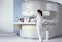
In addition to trying to improve those results with the addition of the 3-point Dixon gradient echo sequence, the group also wanted to test the feasibility of imaging patients on an upright, weight-bearing vertical MR unit, Neumann said.
This experimental study used three cadavers and two normal subjects. The signal-to-noise ratio (SNR) and the contrast-to-noise ratio (CNR) were determined in various tissues. The 0.5-tesla MR imaging protocol on a Signa SP (GE Medical Systems, Waukesha, WI) open scanner for the T1-weighted sagittal spin-echo (SE) images specified TR 500 ms, TE 20 ms, bandwidth 6.25 kHz, variable bandwidth, extended dynamic range, 16 x 16 field-of-view, 4 mm slice thickness, 0 mm skip, matrix 256 x 192, NEX 1.
The three-point Dixon gradient echo images involved TR 150 mm, TE 12.8, bandwidth 15.6 kHz, variable bandwidth, 16 x 16 FOV, 4 mm slice thickness, 0 mm skip, 256 x 192 matrix, NEX 2.
According to the results, the mean SNR on the T1-SE sequence was 14.1 for bone, 11.7 for cartilage, and 12.3 for fluid. The mean CNR between cartilage and bone was 2.4. The mean CNR between cartilage and fluid was 0.6.
On the 3-point Dixon GRE images, the mean SNR was 2.0 for bone, 12.4 for cartilage, and 15.8 for fluid. The mean CNR between cartilage and bone was 8.5. The mean CNR between cartilage and fluid was 2.9. In addition, two of the cadavers had areas of cartilage loss that were accurately depicted using this sequence.
This imaging sequence is particularly attractive when traditional, chemically fat-selective, fat saturation techniques fail, Neumann said. Imaging patients in the upright, weight-bearing position also decreased the cartilage thickness by 15%-20%, making this technique a unique, in-vivo way to assess the biomechanics of cartilage loading, she added.
Other researchers would agree. A team from Nagoya University School of Medicine in Japan stated that the 3-point Dixon sequence might be “the only practical choice for obtaining fat suppressed, T1-weighted images for joint MR.” They used the 3-point Dixon method on a 0.35-tesla scanner for optimizing knee scans (Nagoya Journal of Medical Science, May 2000, Vol.63:1-2, pp. 41-49).
Neumann said her group’s future studies will look at whether the 3-point Dixon GRE protocol is useful for imaging cartilage at varying degrees of knee flexion.
By Shalmali Pal
AuntMinnie.com staff writer
December 2, 2002
Copyright © 2002 AuntMinnie.com

.fFmgij6Hin.png?auto=compress%2Cformat&fit=crop&h=100&q=70&w=100)



.fFmgij6Hin.png?auto=compress%2Cformat&fit=crop&h=167&q=70&w=250)


