Patients who experience a traumatic brain injury (TBI) may also experience long-term psychiatric disorders a year after their head injuries, according to a report in the Archives of General Psychiatry
"The implication for clinicians, in general, is that patients with traumatic brain injury are at high risk for developing major depression with prevalence rates being close to 30% during the first year following TBI," wrote Dr. Robert Robinson, chairman of the department of psychiatry at the University of Iowa in Iowa City.
Robinson, along with co-author Dr. Ricardo Jorge and colleagues, enrolled 91 subjects with TBI in the study. The control group consisted of 27 patients with multiple trauma, but without any evidence of central nervous system injury. Subjects were evaluated at three, six, and 12 months following injury.
At the three-month follow-up, the patients underwent MR on a 1.5-tesla scanner (Signa, GE Healthcare, Waukesha, WI) at the university’s radiology department. Volumetric data was generated using an in-house software program (BRAINS, department of psychiatry, University of Iowa). The software allows cross-modality image registration, automated tissue classification, automated regional identification, cortical surface registration, volume and surface measurements, 3-D visualization of surfaces, and multiplanar telegraphing. Traumatic lesions were classified using the Traumatic Coma Data Bank criteria.
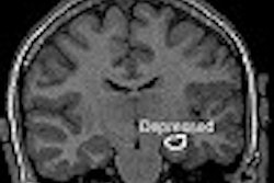
According to the results, the incidence of major depression correlated to reduced gray matter volumes on the brain scans. Thirty of the 91 TBI subjects (33%) suffered from major depressive disorder during the first year after their injury, and the condition was significantly more frequent among TBI subjects than controls. Of the clinically depressed TBI subjects, 76.7% also had anxiety, and 56.7% showed aggressive behavior (Archives of General Psychiatry, January 2004, Vol. 61:1, pp. 42-50).
"Major depression was also associated with poorer social functioning at the six- and 12-month follow-up visits, as well as significantly reduced left prefrontal gray matter volumes, particularly the ventrolateral and dorsolateral regions," Jorge noted.
Previous MR and CT studies conducted soon after acute traumatic brain injury have shown that lesions affecting the left frontal cortex or left basal ganglia are associated with an increased frequency of depression, according to Robinson.
The latest study goes a step further by imaging the patients three months after the trauma, indicating that "the decreased left frontal gray matter volume presumably relates to atrophy following contusion or multiple lesions or shear injury involving the left frontal cortex. A mild degree of left frontal atrophy may indicate that the patient is a significant risk for developing depression over the next year following trauma," he said.
By Bruce SylvesterAuntMinnie.com contributing writer
February 12, 2004
Related Reading
Brain structure may vary in teens with major depressive disorder, February 12, 2004
Echo-planar MR spectroscopy improves mood in depressed patients, January 15, 2004
Prolonged depression reduces global cerebral gray matter volume, November 20, 2003
Functional MRI shows effects of antidepressants, February 28, 2003
Copyright © 2004 AuntMinnie.com


.fFmgij6Hin.png?auto=compress%2Cformat&fit=crop&h=100&q=70&w=100)


.fFmgij6Hin.png?auto=compress%2Cformat&fit=crop&h=167&q=70&w=250)


