Advances in neonatal cardiac surgery have vastly improved newborn survival rates, but caring for these infants is still a delicate undertaking. Impaired neurodevelopment remains a major concern in these high-risk patients, and physicians must also worry about preventing neurological damage.
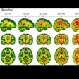
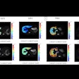
A multidisciplinary group from Philadelphia used pulsed arterial spin-labeled MRI (PASL-pMRI) to assess neonates, and found that preoperative cerebral blood flow (CBF) is diminished in cases of severe congenital heart defects.
Neonates with heart defects frequently suffer from preoperative periventricular leukomalacia (PVL). This condition arises from injury to immature oligodendrocytes, which are prone to ischemia, explained Dr. Daniel Licht and colleagues in the Journal of Thoracic and Cardiovascular Surgery (December 2004, Vol. 128:6, pp. 841-849).
"The significance of PVL in term infants with CHD (congenital heart defects) remains under investigation," the authors wrote. The results of their study "draw attention to the potential role of low CBF being an underrecognized preoperative risk factor for poor neurocognitive outcome."
Licht is from the division of neurology at the Children's Hospital of Philadelphia. His co-author, Dr. Robert Zimmerman, is from the hospital's division of neuroradiology. Other co-investigators are from various departments at the hospital, as well as the Hospital of the University of Pennsylvania, also in Philadelphia.
For this research, 25 term infants (average age 4.4 days) underwent PASL-pMRI on a 1.5-tesla scanner (Magnetom Vision or Sonata, Siemens Medical Solutions, Malvern, PA). Three scans were done immediately before surgery, and were followed by two scans after increased CO2 on a standard ventilator.
Structural MR scans also were obtained during CO2 equilibration. A second pMRI sequence was then done to measure CBF in the hypercarbic state. The images were reviewed for congenital and acquired abnormalities.
The structural MR results showed that 53% of the infants had evidence of developmental and/or acquired lesions, such as PVL, microcephaly, and incomplete closure of the operculum. The mean head circumference was also much less compared to normal neonates. Baseline CBF was 19.7 mL · 100 g-1 · min. Five patients had CBF measurements of less than 10 mL · 100 g-1 · min.
"Significantly, the presence of PVL was associated with low baseline CBF values (p = 0.05) and decreased CO2 reactivity (p = 0.02)," they wrote. "Remarkably, more than 50% of the study cohort had one or more of these (central nervous system) findings that could be attributed to chronic cerebral hypoperfusion."
The group pointed out that preoperative CBF in this patient population was low or even drastically reduced. In addition, low CBF was associated with PVL. While the two repeated perfusion scans after CO2 administration did show high reproducibility, this imaging technique still requires further evaluation. Ideally, a second study with a control group of neonates without congenital heart defects should be performed, the researchers stated.
By Shalmali Pal
AuntMinnie.com staff writer
December 29, 2004
Related Reading
MR may be sounder than sonography for catching fetal abnormalities, November 29, 2004
Neonatal MRI predicts cerebral palsy in neonates at risk, October 15, 2004
Fetal cleft lip and palate seen better with MRI than sonography, July 29, 2004
MRI delineates complexities of preterm infant brain, April 30, 2004
Copyright © 2004 AuntMinnie.com


.fFmgij6Hin.png?auto=compress%2Cformat&fit=crop&h=100&q=70&w=100)


.fFmgij6Hin.png?auto=compress%2Cformat&fit=crop&h=167&q=70&w=250)


