Ovarian carcinoma is a complex disease marked by an overabundance of putative cells of different origins and patterns. The distinction between benign and malignant in this heterogeneous disease has made it difficult to find appropriate early detection methods. Most women are diagnosed at such a late stage that morbidity and mortality rates are high.
Australian MR experts have proposed using in vivo 3-tesla MR spectroscopy (MRS) of the ovary, along with 8.5-tesla MRS of the biopsy specimen, in ovarian cancer. They suggest that the two modalities combined can offer a multitude of information -- preop diagnosis, loss of cellular differentiation, and presence of malignant disease -- for a multifaceted disease.
In previous research, Carolyn Mountford, Ph.D., and colleagues demonstrated that 8.5-tesla MRS on biopsy can reliably distinguish invasive cancer from atypical noninvasive neoplasms, benign neoplasms, and normal tissue (International Journal of Gynecological Cancer, May 1995, Vol. 5:3, pp. 211-221).
Mountford is from the Institute for Magnetic Resonance Research at the University of Sydney. Her co-authors are from other departments at the university, as well as the Royal North Shore Hospital in Sydney.
"The combination of MRI and MRS in vivo ... offers the possibility of preoperative evaluation of complex ovarian masses, which includes malignant and benign lesions, and pelvic inflammatory disease," they wrote in the Journal of Women's Imaging (June 2005, Vol. 7:2, pp. 71-76).
In this case report, the 57-year-old postmenopausal, nulliparous patient had a four-month history of weight loss and abdominal bloating. Physical exam demonstrated a firm abdominal mass arising from the pelvis. An abdomino-pelvic ultrasound revealed bilateral complex masses with ascites.
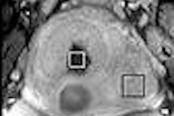
In vivo MR data was collected on a 3-tesla scanner (Magnetom Trio, Siemens Medical Solutions, Erlangen, Germany), and the ovaries were identified on T2-weighted imaging. Two-dimensional chemical-shift imaging (CSI) was undertaken using point-resolved spectroscopy (PRESS). One-dimensional MRS spectra were collected on an 8.5-tesla system (Avance, Bruker BioSpin, Billerica, MA).
The 3-tesla MR and MRS exams showed a mixed cystic and solid lesion measuring 4 cm in diameter. The surgeon used this MR information to obtain biopsies from the ovarian mass intraoperatively. The 2D MR correlated spectroscopy (COSY) exam from the biopsy showed a poorly differentiated serious carcinoma from many organs. On histology, the solid areas of the tumor were predominantly high-grade serous carcinoma with small focal areas of high-grade sarcoma.
The group concluded that this 25 minute exam offers high-quality images, excellent signal-to-noise ratio, and a presurgical modality that will act as an adjunct to current pathologic diagnosis techniques for ovarian serous epithelial tumors.
The COSY spectrum was particularly useful for pinpointing the presence of an adenoma-carcinoma sequence. Finally, noninvasive 8.5-tesla MRS biopsies showed comparable spectral features to those obtained in vivo.
By Shalmali Pal
AuntMinnie.com staff writer
September 14, 2005
Related Reading
MR spectroscopy aids breast MR imaging, July 29, 2005
MR more likely than mammo to expose multiple malignancies in dense breasts, October 13, 2004
MR spectroscopy of biopsy specimen could spare breast surgery, October 1, 2002
Copyright © 2005 AuntMinnie.com


