Fabry's disease is a genetic disorder brought on by the buildup of a particular type of fat in the cells of the body. In its mildest form, it can lead to pain in the extremities, angiokeratomas, hypohidrosis, cloudiness of the cornea, and hearing loss. At its worst, Fabry's disease may lead to life-threatening complications such as kidney damage, heart attack, and stroke.
In the brain and the heart, MRI can track this multisystem disease, offering important information for therapeutic control. While an interdisciplinary group from France laid out the imaging hallmarks of Fabry's disease, researchers in Germany and Italy respectively assessed cardiomyopathy and subcortical ischemic changes.
Fabry imaging features
Most patients with Fabry's disease are successfully treated with enzyme replacement therapy, said Dr. Olivier Lidove and colleagues in their American Journal of Roentgenology pictorial essay. The group is from the departments of internal medicine, radiology, neurology, and nephrology at the Hôpital Bichat Claude-Bernard in Paris.
Imaging results can go a long way toward tailoring that treatment. Lidove's group started out their lecture with the neurological complications of Fabry's disease, most commonly an unpredictable risk for stroke in young men. They advocated MRI for detecting central nervous system (CNS) involvement and for monitoring CNS lesions under enzyme therapy.
On T2-weighted and T2-weighted FLAIR MR studies, readers should look for hyperintense, asymmetric, deep, and widespread white-matter nodules. If the patient is undergoing treatment, these white abnormalities usually remain unchanged, but may worsen if the person is over age 40, the researchers stated. After therapy, the T1-weighted hypersignal of the deep gray-matter nuclei may disappear, they added.
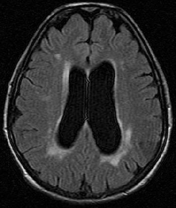 |
| A 52-year-old woman with periventricular hyperintense nodules on FLAIR imaging (1.5-tesla system; TR/TE, 9,000/146; inversion time, 2,250 msec; slice thickness, 5 mm). Nodular pattern, although nonspecific, should suggest disease in nonhypertensive patient and is related to cerebral vasculopathy involving long perforating arteries. Lidove O, Klein K, Lelièvre J, Lavallée P, Serfaty J, Dupuis E, Papo T, Laissy J. "Imaging Features of Fabry Disease" (AJR 2006; 186:1184-1191). |
If the kidneys are involved, cysts can be identified with CT and MR, with the main pattern on the latter being decreased corticomedullary differentiation.
Moving on to the heart, cardiac complications in Fabry's disease may include left ventricular hypertrophy and a short PR interval on ECG. MRI can be employed to diagnose angina pectoris, atrioventricular block, and mitral valve prolapse.
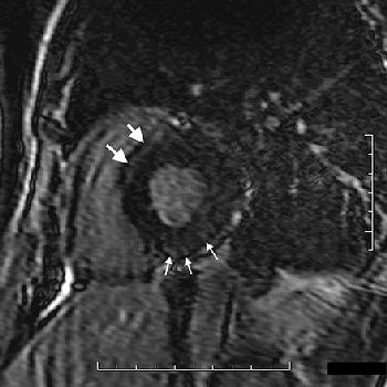 |
| Cardiac involvement in 38-year-old man. Late-enhancement T1-weighted cardiac image in short axis (1.5-tesla system, 3D inversion recovery T1-weighted multishot gradient echo; TR/TE, 3.9/1.4; flip angle, 25°; inversion-recovery prepulse delay, 200 msec) shows band of hyperenhancement assumed to be related to myocardial fibrosis in upper part of septum (large arrows) and subepicardial nodules in inferior wall (small arrows). Cine MRI image at same level (not shown) displayed normal segmental contraction. |
The imaging protocol "must include cine MRI steady-state free-precision sequences in short-axis, long-axis, and four-chamber views; and delayed-enhanced T1-weighted sequences in at least the short-axis view," the authors stated (AJR, April 2006, Vol. 186:4, pp. 1184-1191).
Almost all Fabry's disease patients -- men in particular -- display left ventricular (LV) hypertrophy, brought on by focal myocardial fibrosis, which gives off an abnormal signal. Lidove's group stressed the importance of measuring LV mass at initial presentation as this information must be monitored as a prognostic indicator of therapy, the group said.
Right ventricular hypertrophy
LV hypertrophy is certainly a telltale sing of Fabry-related cardiomyopathy, said Dr. Wolfram Machann in a presentation at the 2006 RSNA meeting in Chicago. His group, from the Bayerischen Julius-Maximilians-Universität in Wuerzburg, Germany, set out to determine if Fabry's disease affected the right ventricle (RV) as well, and if enzyme replacement therapy could stave off RV infection.
While Fabry's disease is quite rare, Machann told AuntMinnie.com that they see more than their fair share of cases at their institution. "We could detect Fabry's disease in 2% to 3% of the patients in our hospital needing hemodialysis due to renal dysfunction," he wrote in an e-mail. "The university hospital of Wuerzburg is a center for the therapy of Fabry's disease. Therefore we see many patients with this rare disease, at the moment 107 patients are under medical attendance in Wuerzburg."
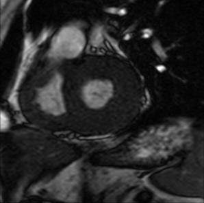 |
| MRI of a patient with left and right ventricular hypertrophy due to Fabry's disease. Image courtesy of Dr. Wolfram Machann. |
For their study, 59 patients with genetically proven Fabry's disease underwent 1.5-tesla MR scans prior to enzyme therapy. Twenty patients were available for follow-up and two healthy controls also underwent MR for comparison.
Their MRI protocol included steady-state-free-precision sequences to measure RV function and mass, as well as cine MRI to determine volume, Machann said. Thirty-nine patients were excluded from the study because of a change in MR protocol from a gradient-echo sequence to a steady-state-free-precession sequence, Machann explained.
For image analysis, the group determined the myocardial mass of the right ventricle, the end diastolic and end systolic volume (from which the stroke volume and ejection fraction were determined). All these parameters were computed in absolute values and normalized to body surface using Argus Software (Varian Medical Systems, Palo Alto, CA).
According to the results, the RV ejection fraction at baseline was 55 ± 6%, which was significantly lower than the control group. During one year of therapy, RV ejection fraction improved to 59 ± 6%. There was also a tendency toward a higher value for the RV mass and a significant difference in systolic and anasytolic volume. His group concluded that RV function can be used as a therapy control parameter in Fabry's disease.
Lidove's group stated that the underlying mechanism for LV hypertrophy in Fabry's disease was focal myocardial fibrosis, which was usually more extensive in men. Machann said that his team did not look for myocardial fibrosis of the right ventricle in this particular project, but they did note a higher RV mass for men because of genetic difference. Men may have more severe disease because women only carry the defect on one chromosome, he added.
Cortical activation
Previous studies have shown that young men with Fabry's disease show multiple lesions in the gray and white matter that is indicative of small-vessel disease. Dr. Cinzia Gavazzi and colleagues from various Italian institutions used brain MR, MR spectroscopy (MRS), and functional MRI (fMRI) to explore the patterns of brain activation in men and women with Fabry's disease.
Gavazzi, as well as co-author Dr. Mario Mascalchi, Ph.D., are from University of Florence. Other contributors are from San Raffaele Hospital and University in Milan and Florence-based Careggi Hospital.
For this prospective evaluation, 16 consecutive patients with Fabry's disease (eight men, eight women) were recruited along with 16 healthy volunteers. Among the Fabry's disease patients, transient ischemic attacks or stroke had occurred in six patients with neurological symptoms. Also, seven patients were receiving enzyme replacement at the time of the study.
All MR exams were done on a 1.5-tesla scanner (Intera, Philips Medical Systems, Andover, MA). For standard MR, the protocol included scout imaging, transverse 3D T1-weighted turbo gradient-echo imaging, transverse T2-weighted fluid-attenuated inversion recovery (FLAIR), and transverse T2*-weighted echo-planar imaging.
For MRS the voxel was placed in the "normal-appearing frontal interhemispheric cortex and subcortical white matter. A point-resolved proton spectroscopy technique was used for acquisition of the proton spectrum," the authors explained. Finally, blood oxygen level-dependent (BOLD) fMRI was done during a 30-second finger-tapping exercise to test motor skills (Radiology, November 2006, Vol. 241:2, pp. 492-500).
The Mann-Whitney U test was used to assess differences in the Fazekas scale (FSS), a ratings system for white-matter signal-intensity changes on FLAIR images, between the following patient pairings:
- Patients versus control subjects
- Healthy men versus men with Fabry's disease
- Healthy women versus women with Fabry's disease
- Men with Fabry's disease versus women with Fabry's disease
- Fabry's disease patients with a history of neurologic disturbances versus those without
- Fabry's disease patients with neurologic deficit versus those without
According to the results, the mean FSS in patients with Fabry's disease was significantly higher than that in healthy controls. Also, the FSS was higher in those patients with Fabry's disease and a history of neurologic disturbances. Additionally, patients with neurologic deficit had a similar FSS to those without.
On MRS, similar results were observed for Fabry's disease patients and controls in terms of N-acetylaspartate/creatine (NAA/Cr), choline/Cr ratios, concentrations of the metabolites, or T2-weighted relaxation times.
Along gender lines, the researchers found no striking difference in FSS between men and women. No significant differences were seen for the normalized brain volume between men and women with Fabry's disease or between male and female controls.
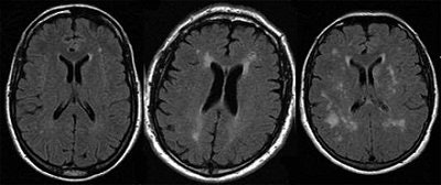 |
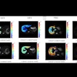
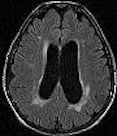
| Transverse FLAIR MR (6000/100/2100) images in patients with FD show examples of varying extent of white matter changes (leukoaraiosis, increased signal intensity) according to the FSS, with a score of 2 in a 36-year-old man (left), a score of 5 in a 52-year-old man (middle), and a score of 6 in a 57-year-old woman (right). Figure 1, Gavazzi C, Borsini W, Guerrini L, et al. Subcortical damage and cortical functional changes in men and women with Fabry's disease. Radiology 2006; 241(2): 492-500. |
"The lack of substantial differences in MR imaging findings between men and women in our study confirms that women should not be viewed as simply carriers of the (Fabry's disease) gene and suggests that they could be enrolled in prospective trials investigating treatments for brain changes associated with this disease," the group wrote.
Based on the intergroup analysis, patients with Fabry's disease showed increased activation of the primary sensorimotor complex bilaterally, the contralateral intraparietal sulcus, and the cingulated motor area bilaterally.
Finally, in patients, there was significant correlation between increased activity in the contralateral sensorimotor cortex and FSS. Overall, the researchers found that subcortical white-matter damage due to small-vessel disease was similar in men and women with Fabry's disease. They also noticed a modification in the cortical activation pattern on fMRI during a motor task. Together, these conditions suggest an adaptive role of cortical functional changes to limit the effects of small-vessel disease, they wrote.
By Shalmali Pal
AuntMinnie.com staff writer
March 9, 2007
Related Reading
Fetal infection with parvovirus B19 may cause neurodevelopmental delays, January 1, 2007
Time-resolved MRA effective for congenital heart defects, October 18, 2006
MDCT complements MR in heart disease evaluation, August 22, 2006
Copyright © 2007 AuntMinnie.com


.fFmgij6Hin.png?auto=compress%2Cformat&fit=crop&h=100&q=70&w=100)


.fFmgij6Hin.png?auto=compress%2Cformat&fit=crop&h=167&q=70&w=250)


