VIENNA - Running a 26-mile marathon can cause both painful and painless ankle pathologies, as anyone who has attempted such a feat can attest. New research presented Saturday at the 2008 European Congress of Radiology (ECR) found that 7-tesla MRI scans can give clinicians insight into just how much ankle damage occurs from endurance running.
Scanning ankles with MRI is a challenge due to their complexity and small anatomical structures. However, in one of the first such uses of a 7-tesla MRI machine, six men were scanned three days after running a full marathon, in the anticipation that the technology's higher spatial resolution would produce clearer depiction of any damage.
Scans were also done with a conventional 1.5-tesla machine within a three-hour time period, and the results were compared to those of the 7-tesla scans. Lead research Dr. Patrick Kokulinsky, of the University of Duisburg-Essen in Germany, had expected to find bone edema, peritendineal fluid, cartilage lesions, and other osseous damage.
"Sports injuries and pain often are not well-correlated. We know from the literature that the mere presence of fluids around tendons, for example, often seems to have no real clinical impact," Kokulinsky said. However, small anatomical structures in the ankle can become wounded by repetitive or intensive exercise.
Comparison of pathologic findings between 7-tesla and 1.5-tesla MRI results revealed that the more powerful magnet provided several advantages.
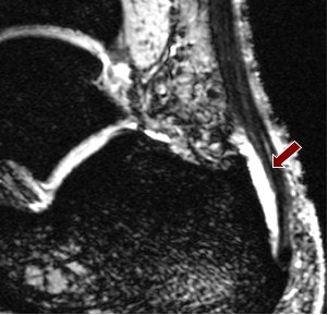
Fluid collection around tendons was apparent in all six runners at both field strengths, though visibility of this was clearer at 7-tesla using a multiple-echo data image combination (MEDIC) sequence, which produced a high contrast with the surrounding tissue.
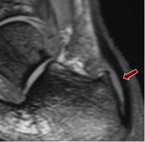 |
| Above, 1.5-tesla image of bursal fluid in the ankle. Below, 7-tesla image shows superior depiction of the bursal fluid. Images courtesy of Dr. Patrick Kokulinsky. |
 |
One runner was identified, at both field strengths, with presumed cartilage defect, yet it was best appreciated with a 7-tesla constructive interference in steady-state (CISS) sequence, due to the sequence's "very high spatial resolution," the researchers said.
Following a marathon, another subject suffered osseous avulsion of the distal tibia, which required the "very high resolution and high contrast" of the 7-tesla magnet to produce the most useful images. Kokulsinky's research found this was best seen using a 7-tesla double-echo steady-state (DESS) sequence.
Surprisingly, there was one instance in which a 1.5-tesla short T1 inversion-recovery (STIR) sequence showed a bone bruise on the anterior compartment of one subject's distal tibia, which was not visible on initial examination of 7-tesla images in any sequence. It was spotted retrospectively, and Kokulinsky was able to explain this inconsistency.
"STIR at 1.5T is particularly helpful for detecting bone bruises, yet for a similarly good contrast at 7T one would choose a high TE (sampling time in the transverse time) for the STIR, but this results in image degradation due to inherently more severe susceptibility artifacts, and therefore a lower TE must be chosen to overcome the problem," he said. "A lower TE (the study used 33 msec), on the other hand, unfortunately results in a decreased T2-weighting, and a lower contrast between fluid and tissue."
This low contrast is most probably the reason why the 7-tesla system produced very poor visibility of the bone bruise, despite the higher spatial resolution seen with 7-tesla STIR sequences.
Both scanners used in the study were manufactured by Siemens Healthcare of Erlangen, Germany (Magnetom Sonata 1.5T and Magnetom 7T).
The overall advantages of 7-tesla MRI come from its higher signal-to-noise ratio, and hence the improved depiction of small pathologies, such as cartilage defects and bone damage, resulting from the higher spatial resolution, Kokulinksy concluded.
"Furthermore, 7T offers new contrasts; for instance, the gradient-echo-based MEDIC sequence offers a very high contrast between fluid and tissue," he said.
Limitations of using the more powerful magnet were that at high specific absorption rate sequences, only two or three slices may be acquired at a time, necessitating multiple acquisitions. Also, the need to use a low TE for STIR sequences represented a compromise. Side-effects related to the magnet's high power were mild and transient.
While just a small study of six patients, the work is notable as it embodies some of the world's first testing of 7-tesla MRI in identifying ankle pathologies. As for whether the technology might find future routine use in the clinical setting, even perhaps initially in professional athletics, Kokulinsky said it was still too early to be definitive.
"We cannot answer that question at the moment, but we are quite convinced that in some cases there might be a benefit of 7T in certain persons in whom 1.5T or 3T does not lead to a conclusive diagnosis," he said. "We can also imagine the impact of 7T in research settings, such as therapy control in cartilage lesions. With the next generation of 7T MRI coils, there will certainly be another big jump in image quality in terms of spatial resolution and homogeneity."
By Rob Skelding
AuntMinnie.com
March 8, 2008
Related Reading
Parallel sequence plus high-field MR improves ankle imaging, June 29, 2007
MRI keeps pace with rapidly evolving musculoskeletal systems of young athletes, May 20, 2007
Studies uphold MRI's role in meniscal tears, propose new sequences, May 16, 2007
Copyright © 2008 AuntMinnie.com


