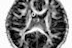
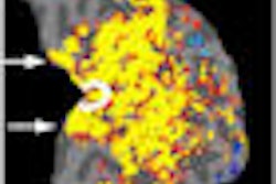
Researchers at the University of Michigan Comprehensive Cancer Center in Ann Arbor are using a new MRI-based analysis method to predict patient survival as early as one week after brain tumor treatment.
The method uses an MRI protocol called a parametric response map to monitor changes over time in tumor blood volume within individual image voxels, rather than a composite view of average change within the tumor.
The researchers studied 44 people with high-grade glioma who were treated with chemotherapy and radiation. Each participant underwent MRI scans before treatment and one week and three weeks after starting treatment.
Looking at standard comparisons using averages, the scans indicated no change one week and three weeks into treatment. However, using the parametric response map approach, researchers were able to show changes in the tumor's blood volume and blood flow after one week that corresponded to the patient's overall survival.
Results of the study appear in the advance online edition of Nature Medicine (April 19, 2009).
Related Reading
CT for brain metastasis unnecessary after PET/CT, April 2, 2009
Diffusion-mapping MRI predicts radiation therapy response, April 22, 2008
Inversion-recovery MRI falls short in brain tumor detection, February 27, 2008
MRI, PET/CT show different strengths in tumor staging, January 26, 2006
Copyright © 2009 AuntMinnie.com


