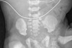
Fetal MRI exams can provide more diagnostic details than ultrasound in assessing fetuses suspected of having neural tube defects, according to a study conducted by researchers in Cairo, Egypt.
The researchers determined that supplemental MRI findings changed or modified the management decisions made with respect to nearly 25% of the fetuses being evaluated. The study was published online in Neuroradiology (June 25, 2009).
Neural tube defects are malformations resulting from failure of the normal neural tube to close early in the embryologic development stage of the fetus. They can include anencephaly, cephalocele, iniencephaly, and spinal bifida. Because the prognosis of the anomaly varies based on its type, size, location, and involvement of neural tissues, prenatal imaging of neural tube defects is of utmost importance.
An ultrasound examination is the primary imaging modality to evaluate a developing fetus suspected of neural tube defects. However, portions of the fetal intracranial anatomy may be obscured, the low sensitivity of fetal sonography to some brain malformations may not show detail, or the maternal body habitus may cause interference.
Dr. Sahar Saleem, a radiologist at Cairo University's Kasr Al-Ainy Hospital, and colleagues conducted the survey to determine the diagnostic utility of MRI exams performed after sonography for revealing suspicious findings, and to determine how the results may have affected parent counseling and pregnancy management.
The researchers retroactively reviewed all records and images from MRI exams performed during a two-year period beginning March 2006. Fetal MRI scans documented neural tube defects in 18 cases and ruled out the ultrasound diagnosis of cephalocele, the herniation of brain matter through a defect in the skull, with the correct diagnosis of oligohydramnios, a condition in which there is too little amniotic fluid around the fetus.
Fetal MRI detected new findings that changed the ultrasound diagnosis in three (15.8%) of the cases, and the modality detected occult findings that caused minor changes in the classification of the anomaly in five (26.3%) cases.
Two pregnancies were terminated as a result of the fetal MRI exam, and one pregnancy that would have been terminated based on ultrasound results alone was continued because of the change in diagnosis. One delivery was changed from vaginal to cesarean. Regarding the majority of the remaining eight cases, additional diagnostic details that benefited the obstetricians were obtained.
Related Reading
MR may be sounder than sonography for catching fetal abnormalities, November 29, 2004
Fetal cleft lip and palate seen better with MRI than sonography, July 29, 2004
MRI second guesses fetal ultrasound, April 25, 2001
Copyright © 2009 AuntMinnie.com


.fFmgij6Hin.png?auto=compress%2Cformat&fit=crop&h=100&q=70&w=100)


.fFmgij6Hin.png?auto=compress%2Cformat&fit=crop&h=167&q=70&w=250)


