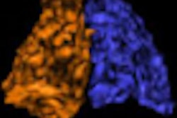
Researchers at Indiana University have detected changes in brain tissue among female breast cancer patients being treated with chemotherapy after surgery, according to a study published in the October issue of Breast Cancer Research and Treatment.
Using 1.5-tesla MRI, the researchers found the most prominent gray matter changes in the areas of the brain that are associated with memory and cognitive functions, such as multitasking and processing speed. On a positive note, the MR images also revealed that gray matter density in most women improved one year after chemotherapy ended.
The lead author of the study was Brenna McDonald, PsyD, assistant professor in the department of radiology and imaging sciences at the Indiana University School of Medicine in Indianapolis (Breast Cancer Res Treat, October 2010, Vol. 123:3, pp. 819-828).
The study is the first to "use a prospective, longitudinal approach to document decreased brain gray matter density shortly after breast cancer chemotherapy and its course of recovery over time," according to the authors. "These gray matter alterations appear primarily related to the effects of chemotherapy, rather than solely reflecting host factors, the cancer disease process, or effects of other cancer treatments."
Prospective enrollment
The study prospectively enrolled 17 female breast cancer patients who were treated with chemotherapy after surgery, 12 female breast cancer patients who received no chemotherapy after surgery, and 18 healthy control subjects. The degree of breast cancer ranged from patients with noninvasive or nonmetastatic invasive disease to those who were treated with standard-dose chemotherapy regimens.
The researchers excluded patients who had received prior treatment with cancer chemotherapy, central nervous system radiation, or intrathecal therapy; had current or past alcohol or drug dependence; or had neurobehavioral risk factors, including neurologic, medical, or psychiatric conditions, that would affect brain structure or function.
All MRI scans were acquired on the same 1.5-tesla system (Signa LX scanner, GE Healthcare, Chalfont St. Giles, U.K.) with EchoSpeed gradients and a standard clinical head coil. The MR images were taken after surgery but before radiation, chemotherapy, and/or antiestrogen treatment to provide baseline results. MRI scans were taken again one month and one year following the completion of chemotherapy.
Gray matter reductions
In the MR images taken at the one-month mark, McDonald and colleagues found gray matter reductions in the bilateral middle frontal gyri and left cerebellum among breast cancer patients who received chemotherapy after surgery. For breast cancer patients with no chemotherapy after surgery, gray matter reductions were found only in the right cerebellum.
By comparison, the authors wrote, there were "no regions where the control group showed lower gray matter than either cancer group at [one month] relative to baseline, nor were there any regions where a significant interaction was found between the two cancer groups from baseline to [one month]."
When MR images were compared from baseline to one-year results, the only significant difference was between breast cancer patients who received chemotherapy after surgery and the control group. The study found gray matter density reductions in bilateral cerebellar regions in patients treated with chemotherapy.
At one month relative to baseline images, breast cancer patients with chemotherapy after surgery also showed decreased gray matter density in bilateral frontal, temporal (including the hippocampus and adjacent medial temporal structures), and cerebellar regions and right thalamus.
Follow-up MR images after one year detected some recovery in those regions, including the bilateral superior frontal, left middle frontal, and right superior temporal and cerebellar regions, although "persistent decreases were also apparent," the authors noted.
Some brain regions demonstrated no recovery over time, where gray matter decreases from baseline to one month were still present after one year, including bilateral cerebellum, right thalamus and medial temporal lobe, left middle frontal gyrus, and right precentral, medial frontal, and superior frontal gyri.
In summary, McDonald and colleagues found breast cancer chemotherapy-related reduction in gray matter density and its partial recovery over time prospectively among female breast cancer patients who received chemotherapy after surgery.
In addition, gray matter changes were most prominent in frontal, temporal, and cerebellar regions, which was consistent with the profile of cognitive dysfunction and complaints observed in certain breast cancer patients after chemotherapy in previous research.
Follow-up research
"This pattern of gray matter changes was not observed in breast cancer patients not treated with chemotherapy [after surgery] or healthy controls, and was not attributable to potential confounds such as disease stage, time since last cancer-related surgery, mood, or medications," the authors added.
The Indiana University researchers are continuing their study to replicate and further investigate the effects of gray matter reduction on the cognitive abilities of cancer patients.
The study was supported by a grant from the Office of Cancer Survivorship of the National Cancer Institute (NCI) and the Indiana Economic Development Corp. (IEDC).
By Wayne Forrest
AuntMinnie.com staff writer
October 11, 2010
Related Reading
MRI helps identify mild cognitive impairment, October 5, 2010
Functional MRI may track aberrant pediatric brain development, September 13, 2010
Study finds diffusion-weighted MRI tops CT in diagnosing stroke, July 12, 2010
MRI hippocampal measurement doesn't predict cognitive decline, April 26, 2010
Resting fMRI exam may predict stroke and brain injury outcomes, March 30, 2010
Copyright © 2010 AuntMinnie.com


.fFmgij6Hin.png?auto=compress%2Cformat&fit=crop&h=100&q=70&w=100)


.fFmgij6Hin.png?auto=compress%2Cformat&fit=crop&h=167&q=70&w=250)


