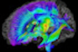
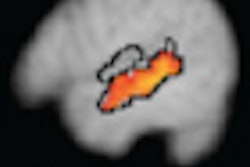
With the help of MRI, researchers at the University of Utah in Salt Lake City have found that areas of the left and right brain hemispheres do not properly communicate with each other in people with autism, according to a paper published online October 15 in Cerebral Cortex.
The study may be an important step in diagnosing autism with MRI, and it could help identify the disorder in children much earlier and lead to improved treatment and outcomes. The research was led by Jeffrey Anderson, MD, PhD, a neuroradiologist and assistant professor of radiology at the University of Utah.
Regions of the brain that showed the lack of communication are associated with functions that are abnormal in people with autism, such as motor skills, attention, facial recognition, and social functioning, according to the study. Anderson and colleagues noted that MR images of people without the disorder did not exhibit the same deficits.
The research included approximately 80 autism patients between the ages of 10 and 35 and took about 18 months to complete. The results will be added to an existing autism study following 100 patients over time.
Janet Lainhart, MD, associate professor of psychiatry and pediatrics and the study's principal investigator, said that the researchers still don't know precisely what's occurring in the brain in autism, adding that the findings contribute an important piece of information to the autism puzzle.
The results add evidence of functional impairment in brain connectivity in autism and provide better understanding of this disorder, she said.
An increasing number of studies have shown abnormalities in connectivity in autism, but the researchers said this study is one of the first of its kind to characterize functional connectivity abnormalities in the entire brain using MRI rather than in a few specific pathways.
The advances highlight MRI as a potential diagnostic tool, so patients could be screened objectively, quickly, and early -- when interventions are most successful. The advances also show the power of MRI to help scientists better understand and potentially better treat autism at all ages.
Anderson and colleagues also hope to use the data to biologically describe different subtypes of autism that may have various symptoms or prognoses, leading to the best treatment for each affected individual.
By Wayne Forrest
AuntMinnie.com staff writer
October 13, 2010
Related Reading
Quick brain scan could screen for autism, August 11, 2010
MEG shows slow response in autistic kids, January 8, 2010
BOLD fMRI may identify the underlying characteristics of autism, November 10, 2009
MRI spots brain abnormality in autistic children, May 4, 2009
Finger tapping with fMRI reveals autism secrets, April 28, 2009
Copyright © 2010 AuntMinnie.com


