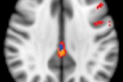
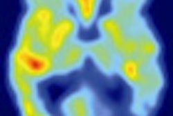
Researchers using arterial spin-labeled (ASL) MRI have found that blood-flow abnormalities can linger or worsen in the brains of veterans with Gulf War illness as long as 20 years after the war ended, according to a new study published online September 13 in Radiology.
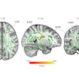
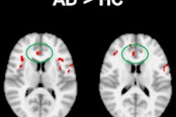
The results showed that abnormal hippocampal blood flow persisted and may have progressed 11 years after initial testing and nearly 20 years after the Gulf War, suggesting chronic alteration of hippocampal blood flow.
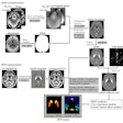
Principal investigator Dr. Robert W. Haley, chief of epidemiology in the departments of internal medicine and clinical sciences at the University of Texas (UT) Southwestern Medical Center in Dallas, and colleagues used ASL-MRI to assess hippocampal regional cerebral blood flow (rCBF) in 13 control participants and 35 patients with Gulf War syndromes.
The hippocampus is responsible for long-term memory function and helps with spatial navigation. The region has been cited as the source of many Gulf War illness-related neurological symptoms, such as memory loss, confusion, irritability, and motion control issues.
Each patient received intravenous infusions of saline in an initial session and physostigmine in a second session 48 hours later. Physostigmine is a cholinesterase inhibitor used to test the functional integrity of the cholinergic system, which is involved in the regulation of memory and learning.
ASL-MRI detected brain abnormalities too subtle for regular MRI, said study co-author Richard Briggs, PhD, professor of radiology at UT Southwestern. The technique allows physicians to make a diagnosis in a single two-hour scanning session without having to expose patients to ionizing radiation.


.fFmgij6Hin.png?auto=compress%2Cformat&fit=crop&h=100&q=70&w=100)

.fFmgij6Hin.png?auto=compress%2Cformat&fit=crop&h=167&q=70&w=250)


